Actor and environmental activist contributes undisclosed amount as part of Israeli company’s recent $105m funding round
Actor and environmental activist Leonardo DiCaprio recently invested an undisclosed amount in Israeli alternative meat startup Aleph Farms, a maker of cultivated meat that grows steaks from modified cattle cells, according to an announcement on Wednesday
The investment was made as part of Aleph Farms’ $105 million Series B funding round in July.
The movie star also backed Netherlands-based alt-meat startup Mosa Meat, according to the announcement. The Dutch company unveiled the first cultured hamburger in 2013 and recently announced an $85 million funding round.
Aleph Farms, meanwhile, rolled out the first cultivated steak in 2018 and a cultivated ribeye cut earlier this year.
DiCaprio will be joining both startups as an advisor, according to the statement. The actor has long championed environmentalism with his eco-focused Leonardo DiCaprio Foundation, giving out $100 million in grants for everything from lion recovery and mangrove restoration to the defense of indigenous rights and better access to affordable solar energy.
In 2019, he joined billionaire investors and philanthropists to create a new nonprofit, Earth Alliance, charged with tackling climate change and the loss of biodiversity.
“One of the most impactful ways to combat the climate crisis is to transform our food system,” DiCaprio said in the statement released on Wednesday. “Mosa Meat and Aleph Farms offer new ways to satisfy the world’s demand for beef, while solving some of the most pressing issues of current industrial beef production. I’m very pleased to join them as an advisor and investor, as they prepare to introduce cultivated beef to consumers.”

Dr. Didier Toubia, co-founder and CEO of Aleph Farms said that “as a committed environmentalist, we welcome Leonardo DiCaprio to our advisory board and family of top-tier investors. Our team is committed to improving the sustainability of our global food systems and we’re thrilled to have Leo share in our vision.”
“With his passion for and dedication to climate action, we expect this collaboration will lead to great things together,” Toubia added in a video announcement.
“Food systems touch all people, and it will take all of us to make this change happen,” he said.
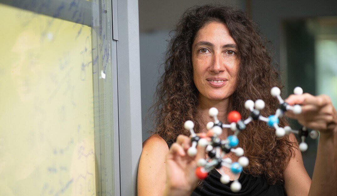
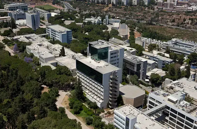
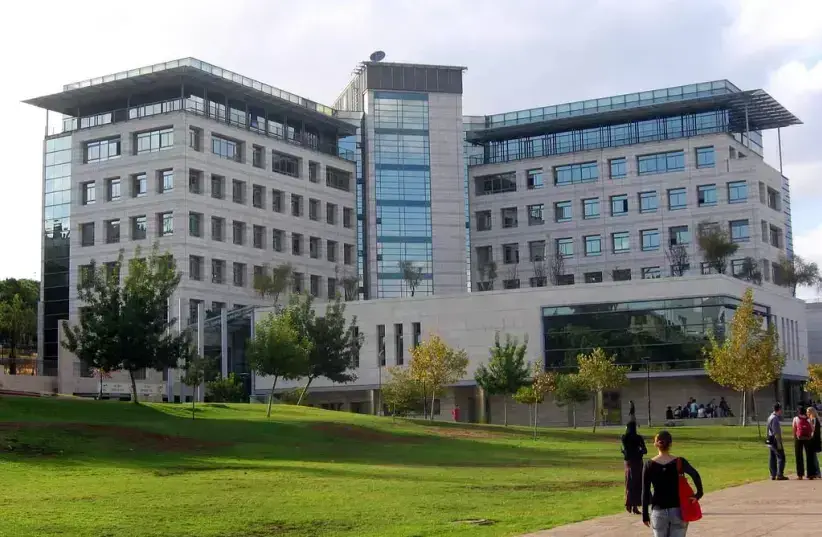
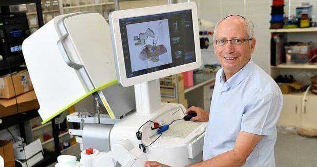
Toubia founded Aleph Farms in 2017 with Professor Shulamit Levenberg of the Biomedical Engineering Faculty at the Technion – Israel Institute of Technology, alongside Israeli food-tech incubator The Kitchen, a part of the Strauss Group.
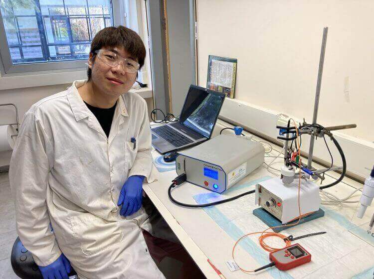
To produce its meat, Aleph leverages the ability of animals to grow tissue muscle constantly and isolates the cells responsible. It then reproduces the optimal conditions for these cells to grow into tissue, basically growing meat outside the animal.

The tissue is grown in tanks that act as fermenters, similar to those in a brewery. There the cells are nurtured and shaped into a 3D structure that makes the meat.
Aleph Farms’ most recent investors include L Catterton, an American-French consumer-focused private equity firm with over $30 billion in equity capital, and DisruptAD, the venture capital arm of the Abu Dhabi holding company ADQ. The startup is also backed by a consortium of global food and meat companies, including Thai Union, BRF, and CJ CheilJedang.
The company has raised more than $110 million to date and has plans for a market launch in 2022. It signed an agreement earlier this year with Mitsubishi Corporation’s Food Industry Group to bring cultivated meat to the Japanese table.

The Israeli firm has also set up similar partnerships with other multinationals: The Swiss industrial group Migros and the United States-based food corporation Cargill have also invested in the startup.
Aleph Farms is a leading player in a growing Israeli food tech sector. The global cultivated meat industry could reach $25 billion by 2030, according to analyst estimates.
Leonardo DiCaprio is investing in Israel’s Aleph Farms
The actor is joining Aleph and Dutch foodtech company Mosa Meat as an investor and adviser in the growing global movement to support sustainable technologies and transform the way meat is produced
Israel’s Aleph Farms and Dutch company Mosa Meat, two foodtech companies in the emerging field of cultivated meat, announced on Wednesday an investment from environmental activist and Academy Award actor, Leonardo DiCaprio. Both companies have demonstrated their ability to grow beef directly from animal cells, with the unveiling of the first cultivated hamburger by Dutch Mosa Meat in 2013 and the first cultivated steak and ribeye by Aleph Farms in 2018 and 2021.
“One of the most impactful ways to combat the climate crisis is to transform our food system. Mosa Meat and Aleph Farms offer new ways to satisfy the world’s demand for beef, while solving some of the most pressing issues of current industrial beef production. I’m very pleased to join them as an adviser and investor as they prepare to introduce cultivated beef to consumers,” DiCaprio said.

With global meat consumption projected to grow between 40%-70% by 2050, cultivated meat offers a solution to reduce negative impacts of industrial beef production, which uses precious sources such as land, water, and also causes harm to animals, while being a leading cause of carbon and nitrogen emissions. Cultivated meat will enable diners to enjoy the qualities of the meat they love, while eliminating the need for cutting out meat altogether. Analysts have projected the cultivated meat market could reach $25 billion by 2030, as part of the broader protein transformation.
According to an independent Life Cycle Analysis study, cultivated beef production is projected to reduce climate impact by 92%, air pollution by 93%, and use 95% less land and 78% less water when compared to industrial beef production.
“As a committed environmentalist, we welcome DiCaprio to our advisory board and family of top tier investors. Our team is committed to improving the sustainability of our global food systems and we’re thrilled to have Leo share in our vision,” Didier Toubia, co-founder and CEO of Aleph Farms said.
Aleph Farms grows beef steaks, from non-genetically engineered cells isolated from a living cow, without harming animals and with a significantly reduced impact to the environment. The company is supported by The Kitchen Hub of the Strauss Group, and Professor Shulamit Levenberg from the Biomedical Engineering Faculty at the Technion – Israel Institute of Technology. Some of its investors include L. Catterton, DisruptAD (ADQ), BRF, Thai Union and Cargill.
Mosa Meat is a global food technology company pioneering a cleaner, kinder way of making real beef. Headquartered in Maastricht, the Netherlands, Mosa Meat is a privately-held company backed by Blue Horizon, M Ventures, Bell Food Group, Nutreco, Mitsubishi Corporation and others.
DiCaprio invests in Israeli cultivated meat co Aleph Farms
The Rehovot-based company has cultivated the world’s first slaughter-free ribeye steak, using 3D bio-printing technology.
Israeli cultivated meat company Aleph Farms has announced that the actor Leonardo DiCaprio has invested in the company and will join its advisory board. DiCaprio is also investing in Dutch cultivated meat company Mosa Meat.
DiCaprio said, “One of the most impactful ways to combat the climate crisis is to transform our food system. Mosa Meat and Aleph Farms offer new ways to satisfy the world’s demand for beef, while solving some of the most pressing issues of current industrial beef production. I’m very pleased to join them as an advisor and investor, as they prepare to introduce cultivated beef to consumers.”
DiCaprio invested in Rehovot-based Aleph Farms as part of its $105 million financing round completed in July. The amount of the investment has not been disclosed.
Aleph Farms cofounder and CEO Didier Toubia said, “As a committed environmentalist, we welcome Leonardo DiCaprio to our advisory board and family of top tier investors. Our team is committed to improving the sustainability of our global food systems and we’re thrilled to have Leo share in our vision.”
Aleph Farms was founded by Israeli food company Strauss Group together with Prof. Shulamit Levenberg of the Faculty of Biomedical Engineering at the Technion – Israel Institute of Technology and Toubia and has cultivated the world’s first slaughter-free ribeye steak, using 3D bio-printing technology and natural building blocks of meat – real cow cells, without genetic engineering.
Leonardo DiCaprio Invests In Israeli Cultivated Meat Startup Aleph Farms

Academy Award-winning actor and environmental activist Leonardo DiCaprio recently invested an undisclosed amount in Israeli startup Aleph Farms, a cultured meat startup that has created slaughter-free steak and ribeye from cattle cells. His investment was part of the company’s $105 million Series B funding round in July, according to an announcement. released on Wednesday.
He also invested in Dutch alternative meat startup Mosa Meat, Aleph Farms and Mosa Meat said in the statement. The Netherlands-based company is known for unveiling the first cultured hamburger in 2013.
Aleph Farms is also known for unveiling the “world’s first” cultivated steak in 2018 and a cultivated ribeye steak earlier this year.
“One of the most impactful ways to combat the climate crisis is to transform our food system,” DiCaprio said in the announcement, “Mosa Meat and Aleph Farms offer new ways to satisfy the world’s demand for beef, while solving some of the most pressing issues of current industrial beef production. I’m very pleased to join them as an advisor and investor, as they prepare to introduce cultivated beef to consumers.”
DiCaprio has a long association with environmental activism and social responsibility, which started early on in his career. In 1998, at the age of just 24, the Oscar-winning actor established the Leonardo DiCaprio Foundation (LDF) with the purpose of raising awareness about environmental issues threatening the health of the planet and to date, has awarded more than $80 million in grants, funding over 200 projects in 50 countries.
In addition, the philanthropist also serves on the board of several environmental protection organizations including the World Wildlife Fund, the Natural Resources Defense Council, International Fund for Animal Welfare, Pristine Seas and Oceans 5. He is also an advisor on The Solutions Project, an organization dedicated to scaling up the adoption of clean, renewable energy.
He also has a history of investing in Israeli eco-friendly projects, including a green hotel at the Herzliya marina, as well as promoting the development of – at the time – in January 2017, the world’s tallest solar thermal tower created by Megalim at the Ashalim solar complex in the Negev.
Aleph Farms has consistently made the news over the last several years as it attempts to disrupt the traditional meat market with its cultured, slaughter-free meat. It grows beef steaks, from non-genetically engineered cells isolated from a living cow, without harming animals and with a significantly reduced impact to the environment.
The company was co-founded in 2017 by Didier Toubia, The Kitchen Hub of the Strauss Group, and Professor Shulamit Levenberg from the Biomedical Engineering Faculty at the Technion – Israel Institute of Technology.
In January, the company announced an agreement with Japanese multinational Mitsubishi Corporation’s Food Industry Group to bring cultivated meat to Japan, followed by a deal to operate in Brazil.