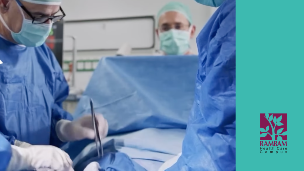
The hospital-developed system analyses medical histories, laboratory results and imaging needs in real time, cutting admission delays and flagging hidden emergencies including a severe infection, suspected child abuse and a life-threatening potassium deficiency.

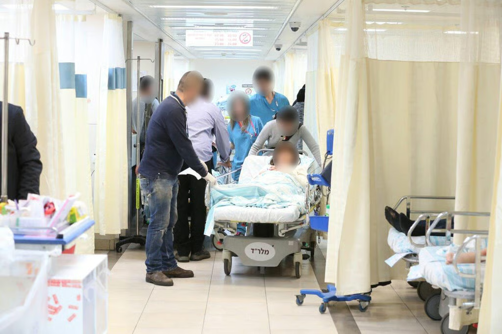
A new artificial intelligence system operating at Rambam Health Care Campus analyses the data of every patient in its emergency departments in real time, suggests possible diagnoses and treatments and identifies complex or unusual cases within seconds.

The system, named Shaked, has been in use at the Haifa hospital for the past six months. According to its developers, it has already shortened the time required to admit patients by one hour and reduced waiting time for specialist consultations in the adult and paediatric emergency departments by 25 minutes.

โ€œThis is the first AI system involved at highly important decision-making points in the emergency department,โ€ said Prof. Shahar Shelly, director of Rambamโ€™s Neurology Department and head of the artificial intelligence laboratory at the Technionโ€™s medical faculty, who initiated and developed the system.

โ€œWe believe waiting times will be shortened significantly in the near future. The results have already amazed us.โ€

Artificial intelligence has assumed a growing role in health care in recent years. Hospitals and health funds are developing and adopting systems that analyze medical information, support clinical decisions and assist physicians in treating patients.

At the same time, the health system remains concerned about doctors independently using external AI products such as ChatGPT, Gemini or Claude, which may not operate within secure medical databases or under institutional oversight.

Shaked supports medical teams throughout the emergency-room process, from admission, diagnosis and treatment to the decision to hospitalize or discharge a patient.

The system consolidates each patientโ€™s medical history, compares it with similar cases, receives and analyzes laboratory results and prioritizes imaging examinations such as CT scans in real time for all patients currently in the emergency department.

โ€œThe system sends alerts to doctorsโ€™ computers about specific patients whose condition could deteriorate within a short period, even when their clinical indicators appeared reasonable during triage,โ€ Shelly said.

It was gum inflammation, not a stroke

Shelly described a recent case in which the system identified severe gum inflammation in a patient whose symptoms initially appeared consistent with a stroke.

โ€œA young woman arrived with neurological symptoms characteristic of a stroke, including paralysis on one side of her face that had developed very rapidly,โ€ he said.

โ€œBecause of the suspected stroke, we were preparing to activate the stroke protocol, including an urgent CT scan. Although neither the patient nor the doctor made the connection, the system alerted us that two weeks earlier she had undergone a complex dental procedure that could become complicated, create a localised infection in the jaw and place pressure on the facial nerve.โ€

(Photo: Elad Gershgoren)

An oral and maxillofacial specialist was summoned to the emergency department and determined that the woman was suffering from acute gum inflammation rather than a stroke.

Another particularly sensitive case occurred in Rambamโ€™s pediatric emergency department after a 4-month-old girl arrived with a fractured thigh.

โ€œThe parents suggested that their 3-year-old child may have fallen on her, but the system retrieved an emergency-room visit from a month earlier involving another injury,โ€ said Dr. Idit Pasternak, director of the pediatric emergency department at Rambamโ€™s Ruth Rappaport Childrenโ€™s Hospital.

โ€œAnalyzing the case and combining the current fracture with the previous injury immediately raised suspicion of child abuse. Without the artificial intelligence, there would certainly have been a consultation with a specialist and a discussion involving the entire team, but in this case the Shaked system raised a red flag over an injury characteristic of abuse.โ€

Because doctors believed the infantโ€™s life could be at risk, they reported the case to the appropriate authorities without further delay.

A third case involved a 13-year-old boy who complained of muscle pain. According to Pasternak, the system analyzed his blood tests and alerted doctors to the possibility of severe hypokalemia combined with metabolic acidosis, a rare and life-threatening condition caused by an acute potassium deficiency.

โ€œWithout immediate treatment, acute heart and respiratory failure can develop within a short time, leading to cardiac arrest and death,โ€ she said. โ€œThe system saved valuable time. The doctors immediately confirmed the diagnosis and began lifesaving treatment.โ€

โ€˜ER lines will become a thing of the pastโ€™

Shelly said one of the principal causes of extended emergency-room stays is the wait for medical imaging, which can sometimes last several hours and create a risk of its own.

The system evaluates the patientโ€™s symptoms, medical background, current clinical indicators and laboratory findings, compares them with existing medical literature and then recommends whether imaging is necessary and which cases should receive the highest priority.

Physicians review every recommendation and retain responsibility for the final decision.

โ€œThe system identifies specific red flags for conditions such as internal bleeding, stroke, pulmonary embolism and others,โ€ Shelly said. โ€œSuch a case will be placed at the top of the list and marked as urgent and requiring immediate action.โ€

In the near future, the system is also expected to help prepare summaries of findings based on radiological interpretations.

Another tool incorporated into Shaked is a chat interface. Instead of opening dozens of previous patient visits to search for a specific piece of information, a physician can question the system about the patientโ€™s file and medical history.

The tool operates using databases developed over decades within the hospital.

โ€œOur AI does not have to go outside the system to mine information, but relies on the most up-to-date medical databases,โ€ Shelly said. โ€œThis makes the teamsโ€™ work more efficient in terms of both time and precision.โ€

โ€œSince we began operating the AI as a quiet adviser in the background, we have spared patients unnecessary CT scans and identified patients who need hospitalization more quickly,โ€ he added.

โ€œThe system can refresh medical information in real time and changes with every result that arrives. That level of dynamism is the first of its kind.โ€

The system was developed with the support of Rambamโ€™s management and through collaboration involving Pasternak, Emergency Department Director Dr. Alexander Strizhevsky, nursing teams and the hospitalโ€™s Digital and Information Technologies Division, headed by Hagar Trau, together with hospital developers Adi Ahituv and Keren Miron.

Dr. Michal Mekel, director of Rambam, said the hospital had placed innovation, and particularly the development of artificial intelligence, high on its list of priorities.

โ€œWe are allocating extensive resources to provide our patients with the best and most advanced treatment in the world,โ€ she said. โ€œWe are entering a new era in medicine.โ€

โ€œWe believe that within a short time, emergency-room lines will become a thing of the past, and we are moving toward a future of precise and rapid medicine that reduces cognitive bias.โ€

In that future, she said, technology will serve as a lifesaving digital safety net, supporting doctors in their decision-making and freeing them to concentrate on what matters most: their human connection with the patient.
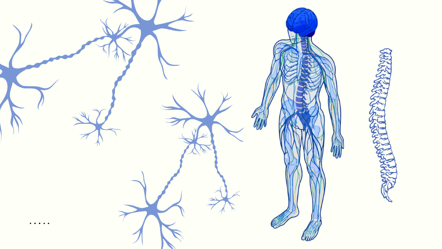
Haifa-based NurExone is preparing for human trials of an experimental therapy that uses engineered exosomes to restore function after spinal cord and optic nerve injuries.

“Our goal is to bring to market a therapy that could help people regain function after spinal cord injuries or restore vision following damage to the optic nerve,” says Dr. Lior Shaltiel, CEO of Haifa-based startup NurExone, which was founded on research conducted at the Technion and Tel Aviv University.

It is an ambitious goal. NurExone is attempting to tackle one of medicine’s most difficult challenges: repairing the central nervous system, where damaged nerves have long been considered largely incapable of regeneration. While doctors have learned to treat many of the complications associated with spinal cord and optic nerve injuries, they have had little success in reversing the damage itself. Patients with severe spinal cord injuries typically do not regain the ability to walk, while damage to the optic nerve often leads to permanent vision loss.

“The moment a spinal cord injury occurs – whether in a car accident or from a partial or complete severing of the cord, for example as the result of a gunshot wound – a secondary injury begins to spread,” explains Dr. Jacob Blumenthal, head of the BrainTech department at the Ehrlich Group, who was involved in the early research conducted at the Technion. “Cells in the affected area begin to die, effectively interrupting communication between the brain and the parts of the body it controls.”

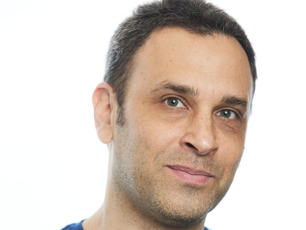
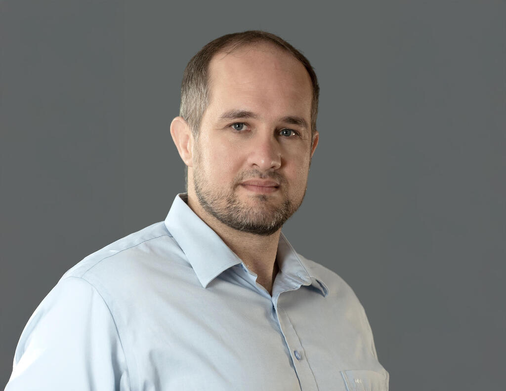
Lior Shaltiel. (Photo: Michal Revivo)

Unlike the peripheral nervous system, where damaged nerves can regenerate, the central nervous system contains biological mechanisms that actively prevent damaged nerve fibers from regrowing.

“If I cut my hand and damage a peripheral nerve, it can regenerate,” explains Yoram Drucker, serial entrepreneur and chairman of NurExone. “But the central nervous system contains molecules that block the rehabilitation of damaged nerves.”

The company’s central question became straightforward: can those biological brakes be temporarily switched off?

Blocking the blocker

One of the key molecules responsible is a protein known as PTEN. Under normal circumstances, PTEN plays a critical role in protecting the body by preventing uncontrolled cell growth, helping suppress cancer. Following a nerve injury, however, the same protein also inhibits the regeneration of damaged nerve cells.

“We know there are inhibitors that prevent nerve growth,” says Drucker. “So the idea was simple: let’s inhibit the inhibitor. If we block the blocker, we allow nerves to regrow.”

The challenge is delivering such treatment directly into damaged nervous tissue without invasive surgery.

NurExone’s approach relies on exosomes, microscopic particles naturally secreted by cells to communicate with one another throughout the body.

“We use exosomes because they can do three things,” Drucker explains. “They naturally migrate toward injured tissue, they help reduce inflammation and promote healing, and they serve as delivery vehicles. We load them with the therapeutic molecule we need, they enter the damaged tissue, release their cargo, and that’s what produces the therapeutic effect.”

The therapeutic cargo consists of a genetically engineered small interfering RNA (siRNA) molecule that temporarily suppresses PTEN activity.

“By temporarily switching off PTEN, we remove the brake on nerve regeneration,” says Shaltiel. “Importantly, we do this only for a short period – roughly two to three weeks after the injury.”

From engineered tissue to exosomes

NurExone’s origins trace back to research led by Prof. Shulamit Levenberg of the Technion and Prof. Daniel Offen of Tel Aviv University, who later became co-founders of the company.

Initially, Levenberg’s team focused on implanting engineered tissue directly into damaged spinal cords.

“We suddenly saw the rats walking,” she recalls. “We expected to see some improvement, but instead they got up, stood on their own, bore weight and walked almost normally. It was an incredibly exciting moment.”

The next challenge was translating those results into a treatment suitable for people.

Performing spinal cord transplants through invasive surgery would be difficult to apply widely, prompting the researchers to ask whether they could use only the biologically active components released by the cells rather than transplanting the cells themselves.

The answer was exosomes.

“We administered only the exosomes through the nose, and again the rats recovered,” says Levenberg. “The animals began walking again. At that point we realized this could become a practical, non-invasive clinical therapy.”

Manufacturing billions of biological delivery vehicles

Producing exosomes at clinical scale presents another technological challenge.

“We grow the cells in bioreactors at 37 degrees Celsius,” explains Dr. Tali Kizhner, NurExone’s vice president of research and development. “The cells grow on tiny carriers inside the bioreactor and continuously secrete exosomes, allowing us to scale production.”

Founded in 2020, NurExone has raised approximately $20 million and is publicly traded on the Toronto Stock Exchange.

Part of the company’s strategy is to pursue regulatory pathways for orphan diseases, which can provide development incentives and potentially accelerate regulatory review.

“This is an extremely active field because there are still no effective treatments,” says Blumenthal. “The first company to successfully commercialize a therapy in this area could gain a significant competitive advantage. Strong patent protection also substantially increases the company’s value.”

The commercial opportunity could also be considerable. Drucker notes that roughly 50,000 new spinal cord injury patients are diagnosed annually across the Western world. The company is also developing treatments targeting optic nerve damage, including glaucoma, which affects an estimated 80 million people worldwide.

NurExone remains in the advanced preclinical stage and is preparing regulatory submissions to the U.S. Food and Drug Administration. The company expects to begin its first human clinical trial in Israel and the United States around 2027, subject to regulatory approval.

But Shaltiel believes the long-term vision extends beyond hospitals.

“Eventually, we’d like to move treatment from the emergency room to the ambulance,” he says. “Someone injured in a car accident could receive the therapy immediately from a paramedic to prevent the loss of nerve cells before they even arrive at the hospital.”

Levenberg shares a similar vision.

“I imagine this becoming available in every emergency department,” she says. “A patient arrives with a spinal cord injury, receives exosome treatment immediately, and we prevent further deterioration while giving the tissue the opportunity to regenerate. If that becomes possible, we could prevent many of the lifelong consequences that spinal cord injuries cause today.”

The vision remains several years away from being tested in humans. But if NurExone’s approach proves successful in clinical trials, it could challenge one of neuroscience’s longest-standing assumptions: that damage to the central nervous system is largely irreversible.

The eye can rebuild lost corneal stem cells by turning old cells back into stem cells, a discovery that could one day help restore vision.

Researchers have found that old, fully mature cells in the cornea can become stem cells again. The cells rebuilt the eyeโ€™s stem cell supply even after every last stem cell had been destroyed.

Cells that had nearly stopped dividing pulled off the reversal and kept the tissue healthy for the rest of the animalโ€™s life.

The cornea is the clear dome at the front of the eye, and losing its stem cells can rob a person of sight.

A repair system the body runs on its own raises the prospect that some blindness could one day be reversed without a transplant from a donor.

A cornea rebuilds itself

The cornea keeps itself clear thanks to a small, hidden workforce. Its own limbal stem cells sit in a narrow ring at the outer rim, called the limbus.

Their offspring travel inward to replace surface cells as they wear away.

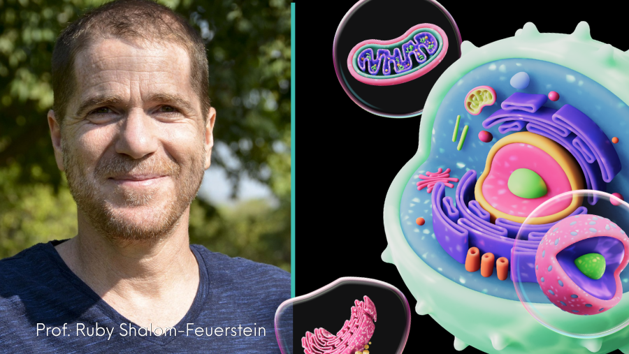
A team led by Professor Ruby Shalom-Feuerstein, a stem cell biologist at the Technion-Israel Institute of Technology, set out to see what happens when that workforce is gone. 

Working with mice, the researchers stripped away the entire ring of stem cells while leaving the surrounding tissue bed untouched. Through the clear cornea, they tracked the repair.

The cornea grew back. Tracing individual cells showed the new stem cells were not survivors that had escaped the scalpel but ordinary mature corneal cells that had reversed course.

Biologists call this dedifferentiation, a process in which a specialized cell reverts to a stem cell.

A remarkable transformation 

The transformation was remarkably complete.

Within about 40 days, the reverted cells were almost indistinguishable from the corneaโ€™s native stem cells, sharing more than 99 percent of their gene activity. Ordinary corneal cells share only 85 to 90 percent.

They also behaved like the real thing, supplying fresh surface cells and keeping the cornea clear for 6 months.

That result ran against a long-held assumption. Losing a tissueโ€™s stem cells was thought to be irreversible, ending in scarring and disease, which is why serious repair has usually meant transplanting cells from elsewhere. 

โ€œWe were surprised to discover that the cornea can regenerate itself even after the destruction of all its stem cells,โ€ said Shalom-Feuerstein.

The oldest cells made a comeback

Cells reversing into stem cells was not new. Earlier work had caught committed cells in fast-renewing tissues doing it.

One influential study showed differentiated cells in the airway becoming stable, working stem cells after the resident ones were removed.

What stayed unclear was the reach of that ability. In those tissues the cells that reverted were young, made only days earlier, so no one knew whether genuinely old cells held the same capacity.

The cornea gave the team a way to test it. Because a corneal cellโ€™s distance from the rim tracks its age, the researchers could pick out the eyeโ€™s oldest cells.

These were mature cells sitting at the center that had spent about four months drifting inward and had all but retired from dividing. Even these old cells made a comeback. 

When moved onto a stripped corneal rim, they switched their stem cell programs back on, rebuilt both the quiet reserve and the active working pool, and kept the cornea clear for nearly a year.

An immune cell trigger

Something had to tell those cells to change. The team traced the signal to macrophages, the immune cells that rush to any injury to clear debris and bacteria. 

They flooded the stripped rim within a day or two, and cutting their numbers by about three-quarters left the stem cells largely unable to return.

The macrophages did more than clean up. As they gathered they released signaling molecules.

Two of them, a growth factor called IGF1 and an immune messenger called CCL2, did most of the work of nudging mature cells back toward a stem cell state.

Blocking the pair stalled the repair, and adding them back restored it.

The reversal has its limits

The reversal has firm limits. It only worked when the tissue bed that houses the stem cells came through the injury intact, and it stayed within the corneaโ€™s own family of cells.

When cells from the conjunctiva, the membrane that covers the white of the eye, moved in to repopulate the corneaโ€™s outer rim instead, they could not transform into corneal stem cells.

The surface clouded and filled with blood vessels โ€“ the signature of blindness caused by stem cell loss.

The eye is not alone. In the skin, researchers have found that the stem cells responsible for hair color can shift in and out of a stem cell state as their surroundings change. 

The finding suggests this kind of cellular flexibility is a normal part of tissue maintenance, not just an emergency response to injury.

A path toward restoring vision

Whether human corneas can do the same is not yet settled. The experiments ran almost entirely in mice, and the team could not directly test reversal in humanย corneal cells.

Working with donated human tissue, they found the same macrophage molecules helped cultured human stem cells keep their identity โ€“ a sign the pathway may carry over.

The stakes are clearest for people who have lost the corneaโ€™s stem cells to burns, infection, or disease. Their vision is already gone or fading.

The study shows that a mature cell, even a very old one, can revert to a fully functional stem cell and remain that way for life, provided the surrounding tissue and immune cells supply the right signals. 

What remains unknown is how to trigger that process on demand and whether other organs possess the same hidden regenerative ability.

โ€œThe next challenge is learning how to control it and how to use it for regenerative medicine,โ€ said Shalom-Feuerstein.

The study is published in the journalย Nature Communications.


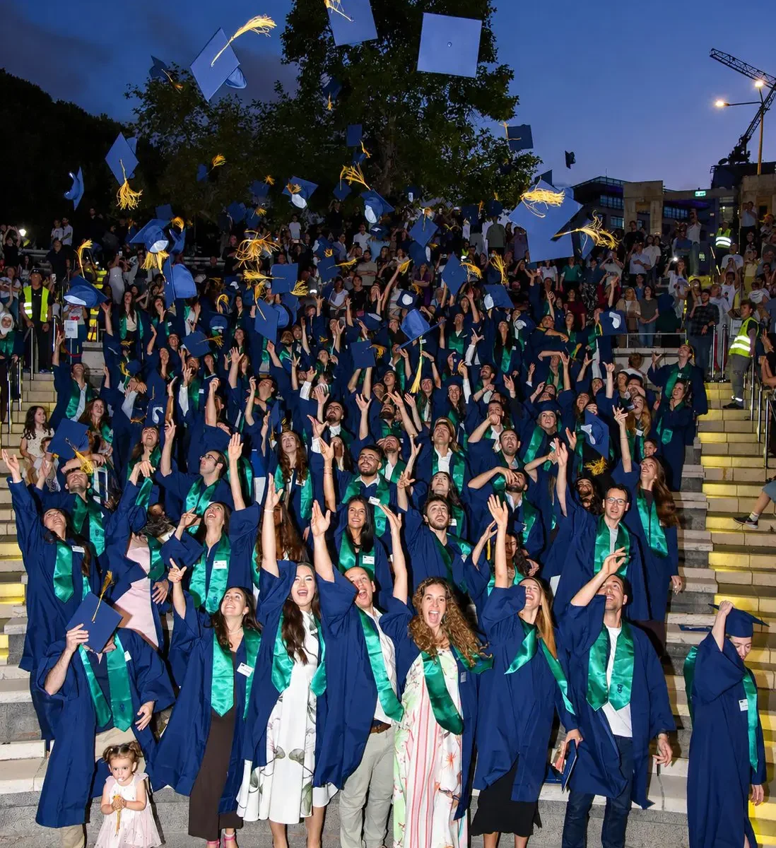
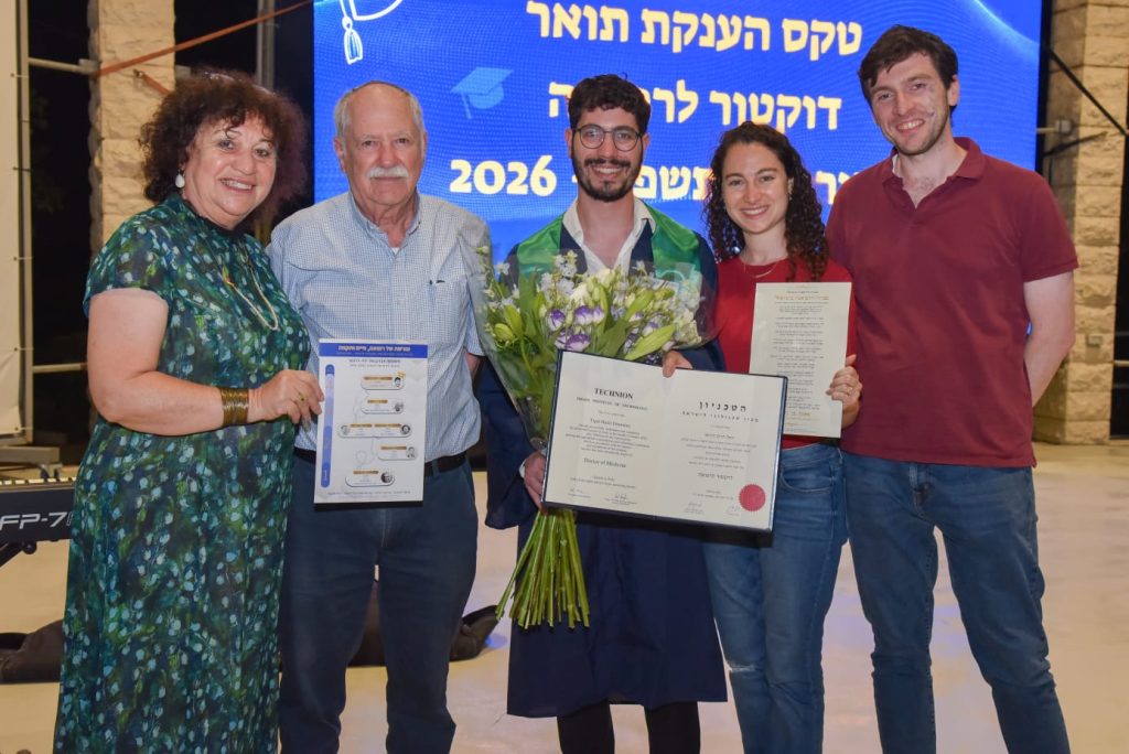
Technion President Prof. Uri Sivan addressed the 118 graduates who completed their medical studies at the Technion Dr. Yigal Haim Drummer, a fourth-generation physician, received his MD degree exactly 100 years after his great-grandfather earned his

The Ruth and Bruce Rappaport Faculty of Medicine recently held its MD graduation ceremony. Attending the event were Technion President Prof. Uri Sivan; Dean of the Ruth and Bruce Rappaport Faculty of Medicine Prof. Ami Aronheim; guest of honor Irit Rappaport; ceremony host and Technion Executive Vice President Prof. Adi Salzberg; faculty members; graduates of the facultyโ€™s 54th graduating class; and their families. A total of 118 graduates received their MD degrees, including 72 women and 46 men. Twenty graduated with honors, eight with distinction, and 12 completed the demanding MD-PhD physician-scientist program.

Addressing the graduates, Prof. Sivan said: โ€œThe profession you have chosen is a truly unique calling โ€“ an intellectual challenge on the one hand, and an expression of generosity, dedication, and compassion on the other. It is a privilege unlike any other, but no less importantly, it is a covenant you enter into today for the rest of your lives. There is no greater privilege and no deeper commitment. The past few years have underscored this beyond any doubt. โ€œThe Ruth and Bruce Rappaport Faculty of Medicine has championed the unique integration of science, medicine, and technology since its establishment more than 50 years ago. Even then, its founders envisioned the future and understood that the future of medicine lay in the close connection between these fields. Today, it is clearer than ever how remarkably farsighted they were.โ€

โ€œYou are individuals with unique stories, backgrounds, values, and dreams,โ€ Irit Rappaport told the graduates. โ€œEach of you has your own passions, talents, and interests, yet you chose this path because you felt called to heal others. I hope that, despite โ€“ and perhaps because of โ€“ the turbulent reality surrounding us and the growing shortage of medical professionals, you will choose to remain in Israel and help shape the future of our children and grandchildren, who, like us, have no other homeland. I wish you every success in all that you do.โ€

Prof. Ami Aronheim told the graduates, โ€œIn addition to meeting the demanding requirements of your degree, you faced extraordinary challenges. The COVID-19 pandemic, a cyberattack on the Technion, and a war that began just two weeks before you were scheduled to start your fifth-year clinical rotations, and that continues to this day. Many of you were called up for hundreds of days of reserve duty. You rose to every challenge. More than that, your class achieved the highest average grades among all six medical schools in Israel.โ€

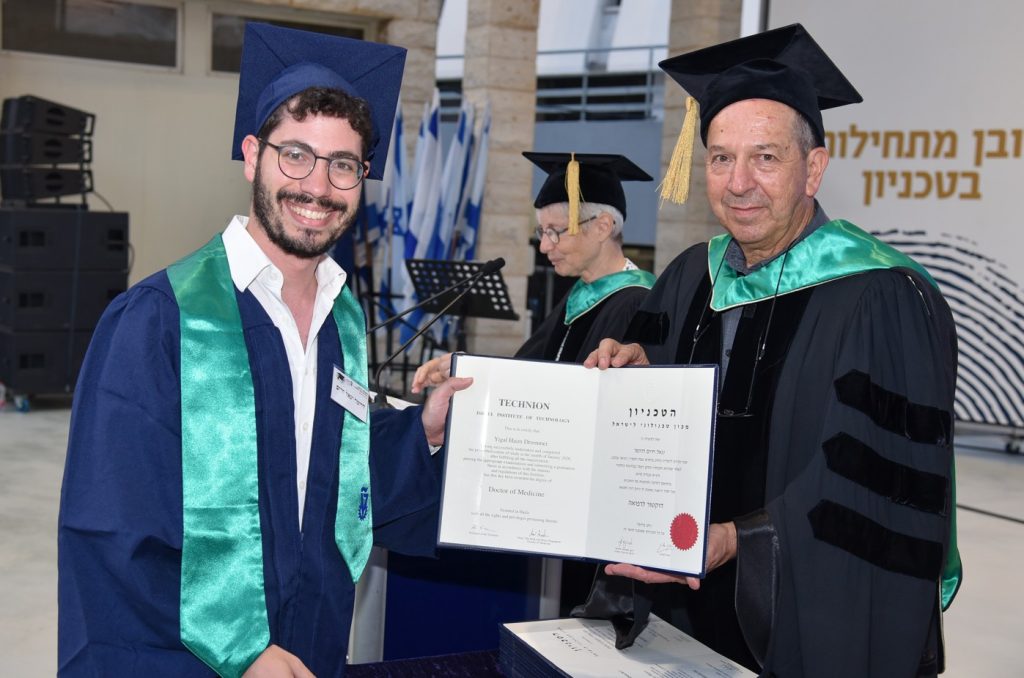
Dean of the Ruth and Bruce Rappaport Faculty of Medicine, Prof. Ami Aronheim, with Dr. Yigal Haim Drummer. Center: Prof. Adi Salzberg

Speaking on behalf of the graduating class, Dr. Liron Krinsky Gonen reflected on their years of study: โ€œThroughout our medical education, we were introduced to the many disciplines that make up the world of medicine, and each of us gradually discovered where our heart truly lies. Wherever we go from here, we leave with an extraordinary set of skills and the knowledge that we studied at the most demanding and professional institution there is. I am certain that each of us will carry the title of graduate of the Ruth and Bruce Rappaport Faculty of Medicine with immense pride, and we will prove it in every department we join.โ€

Four Generations, Six Physicians, One Hundred Years

One of the participants in the MD graduation ceremony was Dr. Yigal Haim Drummer, a fourth-generation physician. Yigal Haim, now 32, grew up in Kiryat Ono and studied at a yeshiva high school, followed by advanced yeshiva studies in Maโ€™ale Adumim. He served in the IDFโ€™s Intelligence Research Division (Lamdan) and was discharged as a captain. He is currently completing his internship at Shaare Zedek Medical Center and plans to specialize in child and adolescent psychiatry.

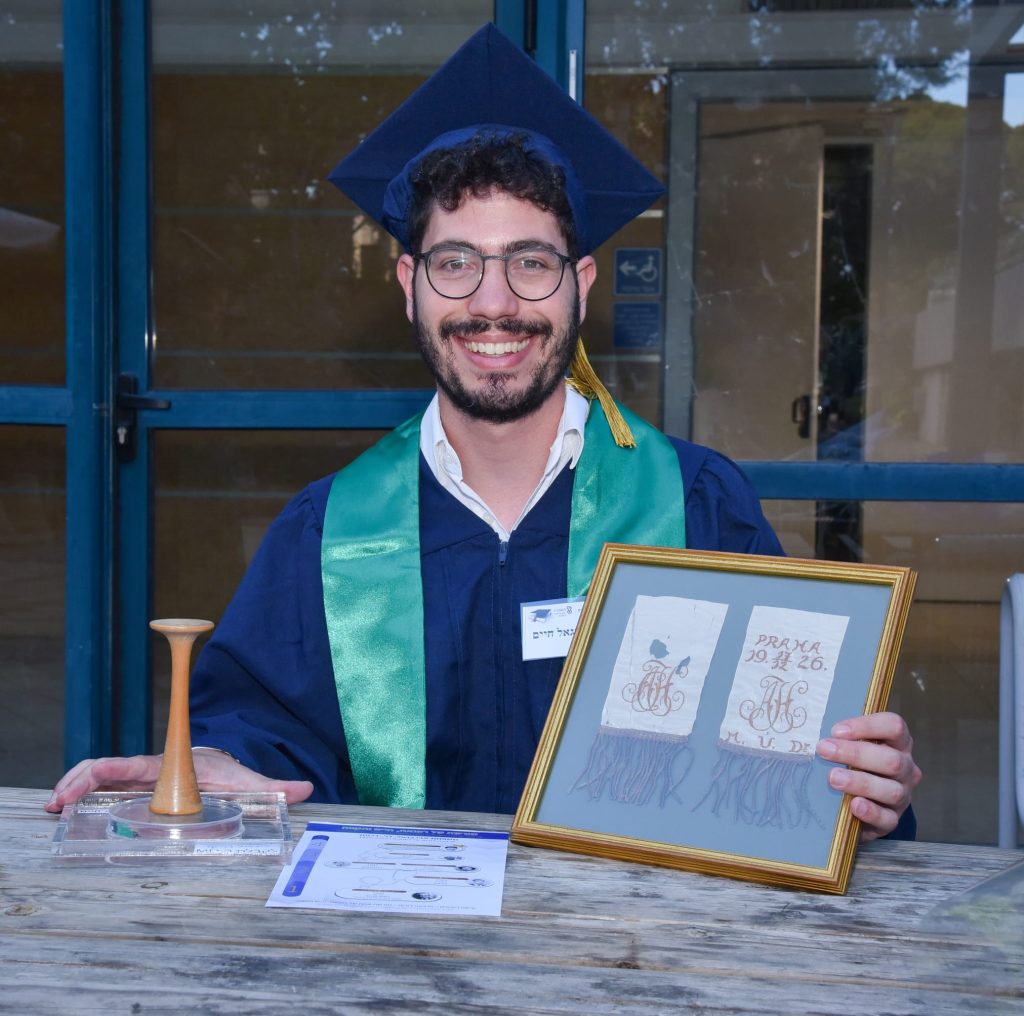
Dr. Drummer is the great-grandson of Dr. Haim Abravanel, after whom he was named. Dr. Abravanel was born in 1896 in Pirot, then part of Serbia. He studied medicine in Prague and Vienna and later directed the hospital in Bitola, North Macedonia. Today, the cityโ€™s day hospital bears his name.

Dr. Abravanelโ€™s children also became physicians. His son, Dr. Nissim Abravanel, was a surgeon in Belgrade, while his daughter, Dr. Reni Levy-Abravanel, was a pediatrician and the wife of radiologist Dr. Salvatore Levy. Nissim, Reni, and Salvatore were killed in the devastating earthquake that struck North Macedonia in 1963.

Reni and Salvatoreโ€™s daughter, Rachel-Shelly Levy Drummer โ€“ who brought her familyโ€™s story to the Technionโ€™s attention โ€“ was orphaned in that tragic event and immigrated to Israel with her grandfather, Haim, and grandmother Berta. In Israel, she married Dr. Dov Drummer, a psychiatrist and psychogeriatric specialist who established the psychogeriatric departments at Kfar Shaul Medical Center in Jerusalem and currently works at Mayanei Hayeshua Medical Center. Rachel-Shelly and Dov are the parents of Dr. Yigal Haim Drummer โ€“ bringing the familyโ€™s medical legacy to four generations.

An interesting discovery by Rachel-Shelly is that exactly 100 years separate the awarding of Dr. Abravanelโ€™s MD degree in Prague in 1926 and the awarding of Dr. Yigal Haim Drummerโ€™s MD degree at the Technion in 2026. Altogether, the family includes four generations and six physicians. Soon, a seventh will join them: Yigal Haimโ€™s sister, Renana, who is currently studying medicine at Tel Aviv University.

ืžืฉืžืืœ: ืจื—ืœ, ื“ื‘, ื™ื’ืืœ, ืื—ื•ืชื• ื•ื‘ืขืœื”
From left to right: Rachel, Dov, Yigal, his sister, and her husband

To mark the completion of his medical studies, Yigal Haim will receive a special gift from his mother, Rachel-Shelly: his great-grandfather Dr. Haim Abravanelโ€™s wooden stethoscope โ€“ a symbol of the human, familial, and professional legacy of the Abravanel, Levy, and Drummer families.

ื™ื’ืืœ ื•ื”ืกื˜ื˜ื•ืกืงื•ืค
Yigal and the stethoscope

Photos: Shay Albaz and Avi Abutbul.

The discovery has potential applications in a wide range of fields, including acoustic cloaking and detection, medical imaging, and underwater communications

Prof. Gal Shmuel from the Technionโ€™s Faculty of Mechanical Engineering is part of a research team that recently won a $7.5 million grant from the U.S. Department of Defense (DoD), led by Prof. Andrea Alรน of the City University of New York.

The Multidisciplinary University Research Initiative (MURI) program is a highly competitive DoD grant program designed to support interdisciplinary teams from multiple universities in conducting groundbreaking basic research that contributes to U.S. national security.

The grant will fund the development of a theoretical discovery made by Prof. Shmuel in 2020, in collaboration with Dr. Pernas-Salomรณn, who was then a postdoctoral fellow under his supervision. This research, supported by the Israel Science Foundation (ISF), wasย publishedย in the leading mechanics journalย Journal of the Mechanics and Physics of Solids, and marked a breakthrough in the field of metamaterials โ€“ engineered materials with properties not found in nature.

Prof. Andrea Alรน. Photo: Paula Vlodkowsky
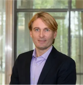
Prof. Gal Shmuel. Photo: Nitzan Zohar

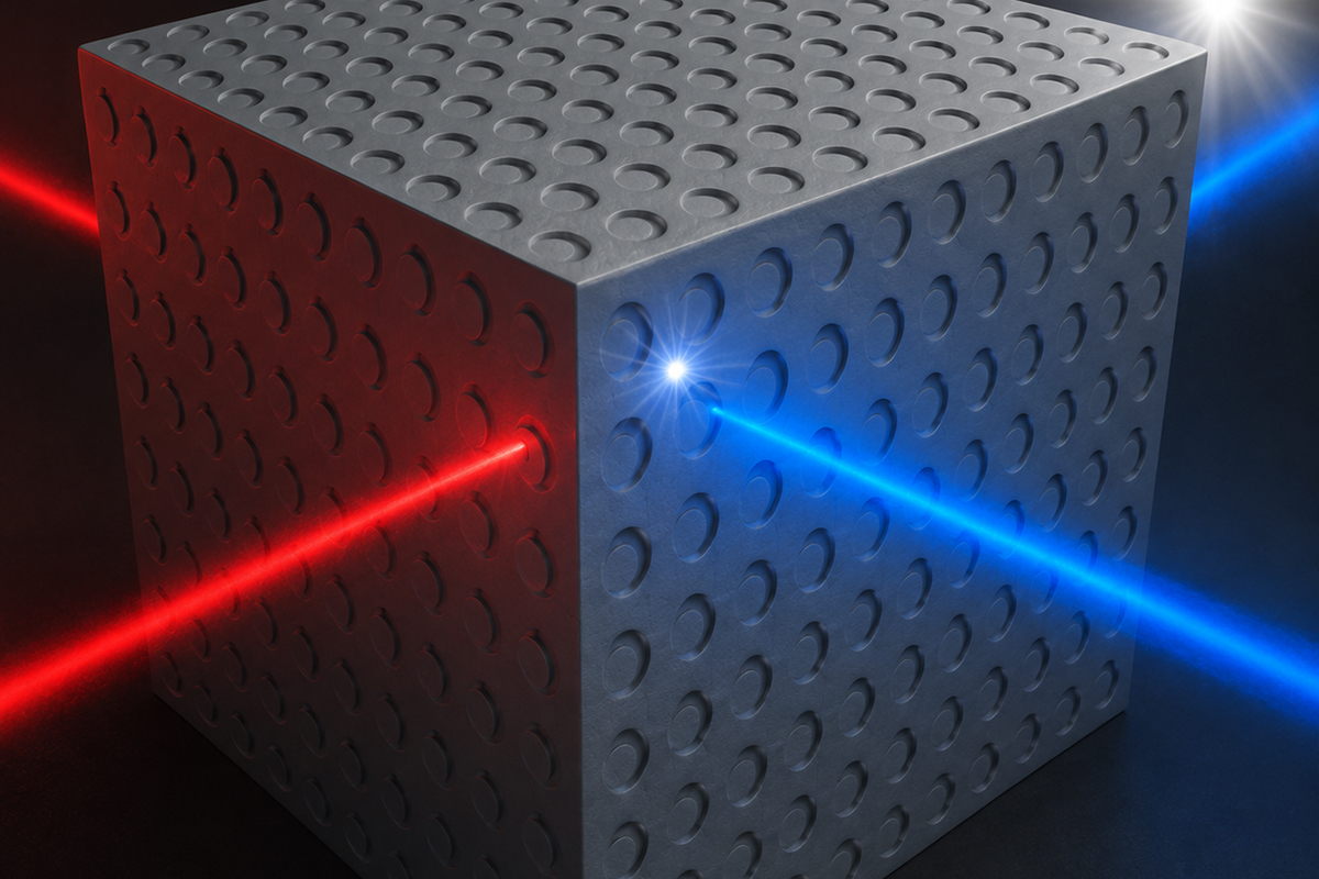
In their paper, the two developed a theory for determining the effective dynamic behavior of electromechanical composite materials โ€“ mixtures of materials whose mechanical and electrical responses are coupled, meaning each depends on the other. According to their theory, by designing such materials in a specific way, the momentum of the composite can be made dependent on the electric field โ€“ a dependency expressed in a unique property that Prof. Shmuel termed the electro-momentum coupling.

The significance of this coupling stems from its role in the balance of momentum in time and space, which is the fundamental physical principle governing the motion of a body and the flow of energy. The electro-momentum coupling designed by the researchers thus offers a controllable degree of freedom for guiding, sensing, and manipulating energy. The coupling has potential applications in a wide range of fields, including acoustic cloaking and detection, medical imaging, and underwater communications.

Following Prof. Shmuelโ€™s theoretical breakthrough, the DoD issued a 2024ย call for researchย aimed at actually creating materials with electro-momentum coupling โ€“ capable of sensing and controlling elastic and acoustic waves via an external electric field.

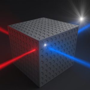
Schematic illustration of an envisioned directional sensor based on electromomentum metamaterials. By engineering asymmetry inside piezoelectric materials, such devices will convert acoustic signals into direction-dependent electrical signals, enabling compact sensing for applications such as acoustic detection, imaging, and underwater communication.

Prof.ย Andrea Alรนย โ€“ one of the worldโ€™s leading researchers in metamaterials โ€“ contacted Prof. Shmuel to jointly draft a research proposal, bringing together five additional researchers from the U.S. and another from Switzerland. The Department of Defense has now announced that the winning proposal is the one submitted by this team, which includes Prof. Shmuel as an international collaborator. In addition, Prof. Shmuel has received a direct grant from the U.S. Army Research Office to support the theoretical and computational component he leads within the broader collaboration.

Not long ago, artificial intelligence felt like something out of science fiction: It lived in futuristic movies and speculative headlines. Today, itโ€™s woven quietly into our daily routines. AI helps us decide where to eat dinner, flags unusual health symptoms, and even drafts our emails. 

But while AI has changed daily life, its impact within research universities may be even more profound. At the Technion, a revolution is unfolding. AI is not just another tool in the academic toolbox. It is transforming how research is done and how quickly discovery happens. 

Technion President Prof. Uri Sivan describes AI as a kind of โ€œsuperbrain,โ€ one we are all connected to. This superbrain can process staggering amounts of information, recognize patterns humans would miss, and solve problems at speeds that were unimaginable just a few years ago. For researchers, whose work depends on thinking, analysing, and discovering, AI has become an extension of their own minds.

A Tectonic Shift in Research 

Across campus, researchers in fields as diverse as medicine, biology, physics, and mechanical engineering are integrating AI into their daily work. Tasks that once required months or even years of painstaking effort can now be completed in hours. Calculations once done by hand or simulations that took weeks to run are now executed almost instantly.

Prof. Mark Silberstein of the Andrew and Erna Viterbi Faculty of Electrical and Computing Engineering believes this transformation is only beginning. โ€œWeโ€™re seeing a tectonic shift in academic research,โ€ he said. โ€œSoon, everyone will be using AI for one thing or another.

AI Revolution | Prof. Mark Silberstein

Within a year, he predicts, AI tools will be fully embedded in research across disciplines, and many researchers will build their own custom AI systems tailored to their work. The pace of change, he said, will only accelerate. 

What does that look like in practice?


From the Test Tube to the Computer

For generations, scientific breakthroughs were born in laboratories filled with microscopes, test tubes, and experimental animals. Today, many of those experiments are beginning not in physical labs, but inside computers. 

Ofer Strichman, professor of computational logic and computer science in the Faculty of Data and Decision Sciences, has watched this evolution firsthand. โ€œEvery year we recruit new faculty,โ€ he explained, โ€œand you can see how more and more scientists are computational experimentalists. Theyโ€™re doing their research in the computer.โ€ 

Prof. Ofer Strichman | AI revolution | American Technion Society
AI revolution | Prof. Ofer Strichman

Imagine developing a new drug. Traditionally, scientists tested one compound at a time, often beginning with animals. Itโ€™s slow, expensive, and limited. Now imagine creating a detailed digital simulation of a human organ, a โ€œvirtual organ,โ€ and testing not just one molecule, but millions of combinations. AI can analyze the results, identify the most promising candidates, and dramatically narrow down what needs to be tested in the lab. Instead of replacing laboratory work, computers supercharge it. Scientists can explore possibilities that would be impossible to test physically, then return to the lab with sharper focus and better odds of success. 

Picture a physicist, for instance, trying to predict how 1,000 celestial bodies will move over the next 1,000 years. The math quickly becomes overwhelming. But with powerful computers, each celestial body can be modeled digitally, with the system calculating how every object influences the others. The simulation unfolds in virtual space, revealing patterns no human could calculate by hand. 

โ€œNowadays,โ€ Strichman said, โ€œthe more computing power you have, the better your research results will be. Like having a bigger telescope, computers allow us to see farther.โ€


Why Computing Power Matters 

Behind every AI breakthrough lies a less glamorous but absolutely essential ingredient: computing power. 

For more than 30 years, the Technion has operated a high-performance computing (HPC) facility: essentially a warehouse filled with powerful servers. These systems have long supported researchers running complex simulations, particularly in fields like physics and engineering.

AI revolution | HPC Building at the Technion
3D render of High Performance Computing Building on Technion campus in Haifa

Traditionally, these computers relied on components called central processing units, or CPUs. You can think of a CPU as the brain of a computer. The Technion currently operates about 6,500 CPUs, and researchers typically wait just a couple of minutes to access one. But AI demands something different. 

Modern AI systems rely heavily on graphics processing units, or GPUs. Originally designed to render video game graphics, GPUs are uniquely suited for the kind of massive, parallel calculations that AI requires. While a CPU handles tasks sequentially, a GPU can perform many calculations simultaneously, making it dramatically faster for AI workloads. The difference is enormous. 

GPUs are not only expensive (each unit can cost around $250,000) but they also require specialized infrastructure. They consume large amounts of electricity and generate extraordinary heat, demanding sophisticated cooling systems and advanced networking to allow thousands of units to communicate seamlessly. The Technion currently has only 72 GPUs, which is far from sufficient. Researchers can wait four hours or more for access to one. In a world where speed determines competitiveness, those hours matter.


A Global Race 

Around the globe, countries, universities, and technology companies are racing to dominate the AI frontier. Success depends not only on talent and ideas, but also on infrastructure. The institutions that build the most advanced computing systems gain a powerful edge in research, innovation, and economic development. 

โ€œThere is an arms race among countries and universities to achieve AI dominance. To be at the forefront of this field, we need to strengthen the capabilities we have at the Technion.โ€

Prof. Mark Silberstein

At present, many Technion researchers must rely on industry partnerships to access advanced GPU systems because the University lacks sufficient in-house capacity. While collaboration with industry can be valuable, dependence creates limitations. 

Complicating matters, Israelโ€™s recent war with Hamas forced national and institutional priorities to shift and long-term infrastructure investments were necessarily delayed. Now, as the country looks toward rebuilding and strengthening its future, expanding AI infrastructure has become a strategic priority. 

The Technion is taking a major step forward with the construction of the Martin and Grace Druan Rosman High-Performance Computer Data Centre. The facility is nearing completion and will provide a state-of-the-art home for next-generation computing. 

Supported by Dr. Martin Rosman and Grace Druan Rosman through the American Technion Society, the new centre includes advanced electrical systems, cutting-edge cooling technologies, and high-speed communications networks โ€” all designed specifically to support powerful GPU-based systems. In simple terms: The building will be ready for the AI era. 

martin and grace rosman unveiling new supercomuting center at the technion in haifa | Donate to Support Israel | Technion University
Martin and Grace Rosman unveiling the supercomputing centre, 2023

High Stakes for Israel 

For Israel, the implications extend far beyond campus. Israelโ€™s reputation as the Startup Nation rests heavily on the strength of its scientific institutions. Many of the countryโ€™s most successful technology companies trace their roots to Technion labs and classrooms. The engineers and entrepreneurs trained here help power Israelโ€™s economy. 

If the Technion falls behind in AI research infrastructure, the ripple effects could be significant. Conversely, if it leads, the impact could be transformative: accelerating medical breakthroughs, advancing clean energy solutions, strengthening national security, and fueling new industries. 

โ€œThe Technion is committed to educating the best engineers in the world, the most capable entrepreneurs,โ€ Silberstein said. โ€œIsraelโ€™s brainpower is our competitive advantage.โ€ 

The AI revolution is here and itโ€™s reshaping science, education, and industry. At the Technion, the question is not whether AI will transform research because that transformation is already underway. The question is how boldly and how quickly the University can build the infrastructure needed to lead.  

Technion scientists

Technion researchers and partners at Tianjin University say managing carbon dioxide, rather than eliminating it, could make low-cost fuel cells more durable and practical for vehicles, drones and remote power systems

Researchers at the Technion-Israel Institute of Technology and Tianjin University in China say they have developed a new approach that could make hydrogen fuel cells more affordable, durable and efficient while allowing them to operate with ambient air.

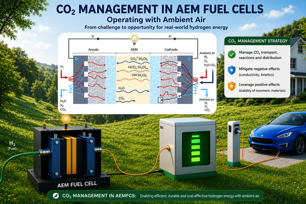
Their findings, published in Nature Energy, focus on anion-exchange membrane fuel cells, or AEMFCs, which generate electricity through a reaction between hydrogen and oxygen. Unlike some conventional fuel cell technologies, AEMFCs can use cheaper and more abundant materials, potentially reducing system costs.

The technology is being studied for use in transportation, aviation, aerospace, drones, distributed energy systems, backup power and electricity generation in remote areas.

The study was led by Prof. Dario Dekel of the Technionโ€™s Wolfson Faculty of Chemical Engineering and the Nancy and Stephen Grand Technion Energy Program; Prof. Michael Guiver, a polymer membrane expert at Tianjin University; Dr. Karam Yassin, manager of the Technionโ€™s Central Hydrogen Technologies Laboratory; and Dr. Sapir Willdorf-Cohen, a researcher in Dekelโ€™s group.

The main challenges in developing AEMFCs have been improving power output, energy efficiency, performance and durability.

Until now, carbon dioxide in ambient air has largely been treated as a contaminant that harms performance and shortens fuel cell durability. The researchers propose what they call โ€œCO2 management,โ€ arguing that carbon dioxide should not be viewed only as an obstacle but as a factor that can be controlled and, under some conditions, used to improve fuel cell stability.

โ€œFor years, carbon dioxide has been considered one of the main challenges facing AEM fuel cells,โ€ Dekel said. โ€œOur work shows that the picture is more nuanced. Under certain conditions, carbon dioxide may contribute to the long-term stability of fuel cell materials. By learning how to manage CO2 rather than simply eliminate it, we can pave the way toward affordable, durable and high-performance fuel cells capable of operating directly with ambient air.โ€

The researchers said the findings could help speed the adoption of hydrogen fuel cells in passenger vehicles, trains, drones, ships, distributed energy systems and autonomous power technologies.

The research was supported by the Nancy and Stephen Grand Technion Energy Program, the Israel Science Foundation, the Israeli Council for Higher Education and other funding partners.

Technion-led study combines deep learning and mathematical modelling to produce dynamic MRI images at up to one frame per second.

A group of researchers from the Technion in Israel and the United States has reported a breakthrough in MRI scanning that could significantly improve breast cancer diagnosis, according to a paper published in Nature Communications.

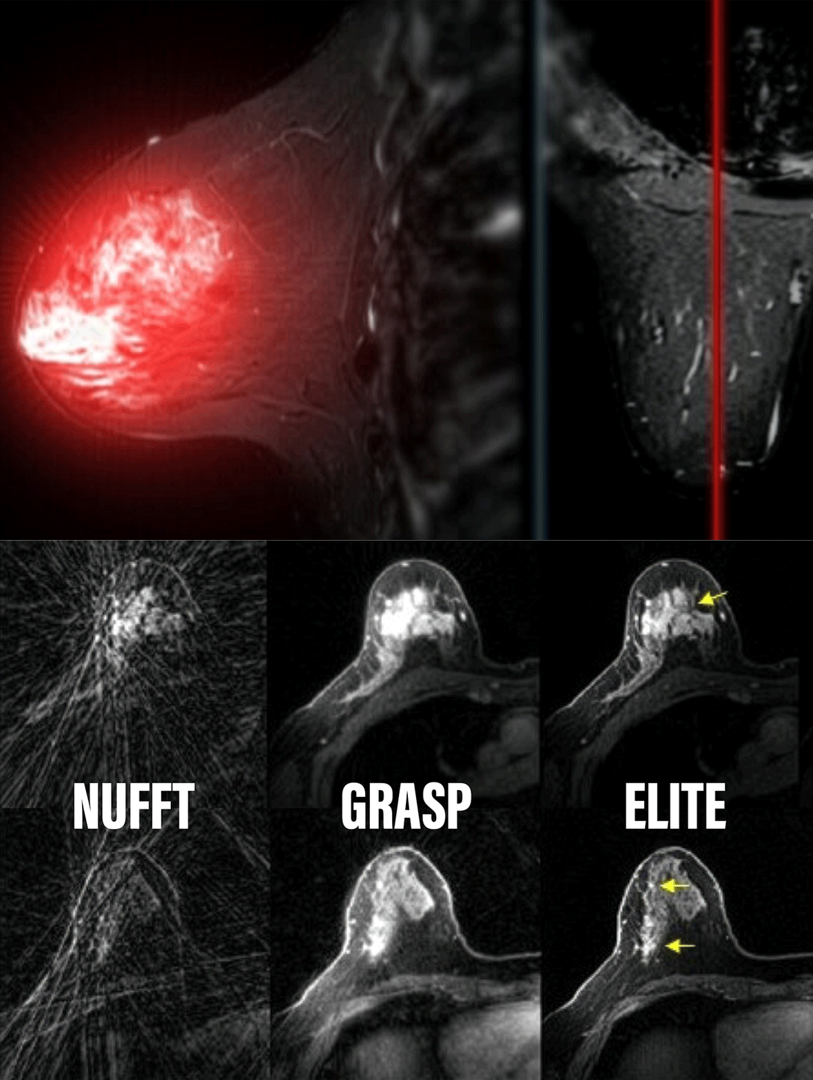
The researchers developed a new method, called ELITE, that accelerates and enhances MRI scans used in breast cancer imaging, a disease diagnosed in approximately 2.3 million people each year, most of them women. The approach combines artificial intelligence with advanced mathematical modelling to enable dynamic MRI imaging at what the researchers describe as unprecedented speed and accuracy.

 Dr. Eddy Solomon
Dr. Eddy Solomon.ย (Leo DeLuca)

The international study brings together expertise in engineering, MRI physics, artificial intelligence and clinical radiology.

Dr. Eddy Solomon of the Technionโ€™s Faculty of Biomedical Engineering, the studyโ€™s lead author, said the research focuses on dynamic MRI, a key tool in breast cancer diagnosis. Dynamic MRI is primarily used for screening high-risk populations and is characterised by high sensitivity, with more than 90% accuracy, compared with roughly 50-60% for ultrasound and mammography combined.

However, MRI technology has long faced a fundamental limitation: producing highly detailed images requires relatively long scan times, making it difficult to track the movement of contrast material through tissue in real time. Traditional MRI systems typically generate one image every one to two minutes at best, limiting the ability to capture the rapid dynamics of contrast agents.

Dr. Solomon and his colleagues sought to bridge this gap by combining mathematical modeling that identifies structural and functional patterns in different tissues with a deep neural network (ResNet) trained to remove noise and distortions. The system also reconstructs missing information from undersampled measurements.

The result, according to the researchers, is the ability to generate one image per second.

The improved temporal resolution allows clinicians to track the movement of contrast agents almost continuously. This, the researchers say, could improve the detection of small tumours, help distinguish more accurately between benign and malignant growths, and better characterise tumour biology, including blood flow and vascular permeability.

In a study involving 54 patients, the researchers reported improved tumour visibility compared with existing methods, higher image quality, and strong diagnostic sensitivity.

They also said that shorter scan times could increase the number of patients that can be scanned using a given MRI system, potentially improving access to imaging services.

The findings are presented as a step toward faster and more precise MRI-based cancer diagnostics, though further validation and clinical deployment would be required before broader adoption.

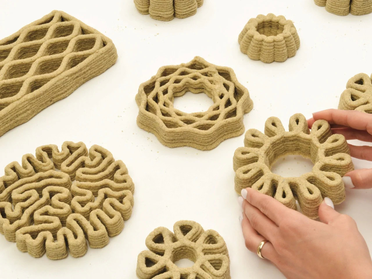
Concrete shapes the world you live in, but its environmental toll is massive.ย Imagine, though, if the buildings around you could grow and breathe, helping to heal theย planet. This isnโ€™t science fiction. Itโ€™s the vision of CyanoCement. Developed by researchers from the Technion,ย Israelย Institute of Technology, this innovative biocement uses ancientย microbesย to redefine how we think about construction materials.

At the heart of CyanoCement are cyanobacteria, tiny photosynthetic organisms responsible for Earthโ€™s first oxygen-rich atmosphere. By leveraging these extraordinary capabilities, the teamโ€”Perla Armaly, Yuval Berger, Lubov Iliassafov, Keren Rosenblau, Yechezkel Kashi, and Shany Barathโ€”crafted a process where these microbes bind minerals and precipitate calcium carbonate, creating a solid without high emissions.

Petri dish with cyanobacteria cultures on a laboratory table.

Innovative Design Meets Environmental Responsibility

This biocement doesnโ€™t just end its environmental work once installed. It continues to capture carbon dioxide from the air, actively working against the problem of atmospheric carbon. Unlike conventional methods, CyanoCement turns construction into part of the solution.

Construction worker applying biocement to a brick wall surface.

The material is designed for facades,ย interiorย panels, and decorative structures. By focusing on non-load-bearing elements, the team keeps the projectโ€™s ambitions grounded, managing expectations with scientific precision.

Close-up of biocement texture with visible green cyanobacteria.

Visible Green: A Living, Breathing Material

The green hue of CyanoCement isnโ€™t painted on. Itโ€™s the color of lifeโ€”indicative of the cyanobacteria within. The design makes environmental benefits visible, offering a reassuring sign that sustainability is working, right before your eyes.

Lab technician measuring pH level of biocement solution.

This innovative project emerged from the Disrupt Design Lab at Technion, in collaboration with the Applied Genomics Lab, marking a significant crossover between architecture and biology. Itโ€™s a fusion rarely seen, yet wholly necessary for the future of sustainable design.

For a unique blend of nature and architecture, explore how theย Sofia Pavilion blends urban landscapeswith natural elements.

Construction site using eco-friendly biocement blocks in foundation.

CyanoCement was honored with the Green Productย Award, celebrated for its meaningful impact and robust research. Itโ€™s not just talkโ€”this material has substance and intention.

Learn how ancient materials are making a comeback withย Finnish designers crafting fashionย from shipwreck timber.

Green biocement samples displayed on a laboratory workbench.

As we think about the future of architecture and sustainability, itโ€™s time to reconsider the role of construction. CyanoCement poses a radical idea: buildings that are not only structures but contributors to the atmosphere. An idea thatโ€™s difficult to ignore once it takes root in your mind.

Researcher writing down observations of growing cyanobacteria samples.

Large language models are an innovative tool transforming a wide range of tasks, including translation, text comprehension, and code generation. However, these models also have shortcomings that require improvement, including biases, disregard for instructions, and โ€œhallucinationsโ€ (i.e. the generation of inaccurate information).

These challenges are a major focus of the research group led by Dr. Haggai Maron from the Andrew and Erna Viterbi Faculty of Electrical and Computer Engineering at the Technion, in collaboration with researchers from other universities and NVIDIA. Recently, three papers by the group were accepted to the most prestigious conferences in computational learning: ICLR 2026, NeurIPS 2025, and AAAI 2026. The papers were led by Ph.D. student Guy Bar-Shalom (co-advised by Prof. Ran El-Yaniv) and postdoctoral researcher Dr. Fabrizio Frasca, in collaboration with Dr. Yftah Ziser (University of Groningen and NVIDIA).

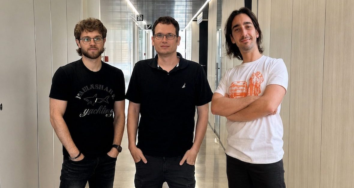
ืžื™ืžื™ืŸ ืœืฉืžืืœ: ื“"ืจ ืคื‘ืจื™ืฆื™ื• ืคืจืกืงื”, ื“"ืจ ื—ื’ื™ ืžืจื•ืŸ ื•ื’ื™ื ื‘ืจ ืฉืœื•ื
In the photo, from left to right: Guy Bar-Shalom, Dr. Haggai Maron, Fabrizio Frasca

Dr. Maron and his team propose a new research direction for identifying failures and flaws in text generated by large language models. Instead of attempting to fully understand how the model operates at every level (something that remains beyond the current reach of the research community), the authors suggest a more pragmatic, inexpensive, and faster approach. Their method is based on building and deploying new machine-learning systems on top of the modelsโ€™ internal computations, in a way that leverages the complex internal structure of those computations. The goal is for these learning systems to detect and utilize hidden information embedded within these computations, even if humans do not fully understand it. The key achievement is demonstrating the possibility of externally and inexpensively monitoring and diagnosing risks. This approach enables users to supervise the model, predict its behavior, and control it without fully understanding the entire mechanism.

The research addresses one of the most critical challenges of the AI era: how to identify when a large language model is making mistakes, fabricating information, or deviating from expected behavior. The methods developed at the Technion provide rapid and effective diagnostics that do not depend on understanding the entire mechanism or the modelโ€™s training process.

The new approach opens broad practical possibilities, including the development of warning systems, quality assurance tools, and safety standards for language models used in medicine, research, education, regulation, and other fields. This marks an important step toward the responsible integration of artificial intelligence into critical systems and toward making AI tools more reliable.

This series of studies is part of a broader research program in Dr. Maronโ€™s laboratory, where the group investigates how patterns can be learned from new types of data that can be extracted from trained models, such as their weights and signals used during training.