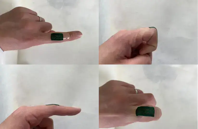
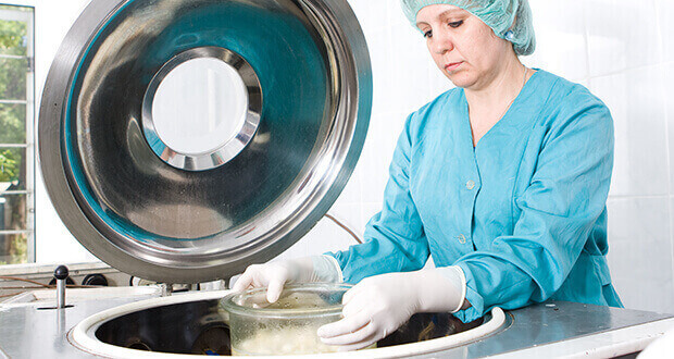
Researchers at the Technion have developed a highly stretchable electronic material and a wearable sensor capable of identifying precise bending and twisting motions.
Scientists at the Technion-Israel Institute of Technology have produced a highly stretchable electronic material and a wearable sensor capable of identifying precise bending and twisting motions.
Essentially, it is an electronic skin.
The development will be able to help identify ailments and disease, for example, the early onset of Parkinson’s, or help amputees adapt to prosthetics, the developers have said.
It recognizes the range of movements that human joints normally makes with the precision of up to half a degree. This breakthrough is the result of collaborative work, headed by Professor Hossam Haick from the Wolfson Faculty of Chemical Engineering.
It was recently published in Advanced Materials, a peer-reviewed journal.
Professor Haick’s lab focuses on wearable devices. Wearable motion sensors can currently recognize bending movement, but not twisting. Sensors that recognize twisting are large and cumbersome.
Ph.D candidate Yehu David Horev and postdoctoral fellow Dr. Arnab Maity have found a way to overcome this problem. Horev found a way to form a composite material that is both usable as a sensor and is flexible, stretchable, breathable, biocompatible, and does not change its electrical properties when stretched.
Dr. Maity was able to solve the mathematics of analysing the received signal.
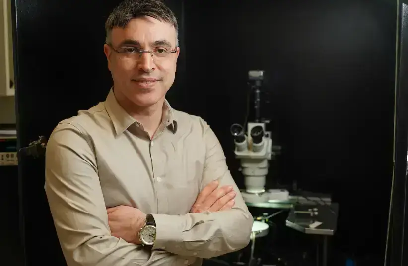
Professor Hossam Haick (credit: TECHNION SPOKESPERSON’S OFFICE)
The novel sensor is breathable, durable and lightweight, allowing it to be worn by humans for long periods of time.
“This sensor has many possible applications,” Prof. Haick stated.
“It can be used in early disease diagnosis, alerting of breathing alterations, and motor system disorders such as Parkinson’s disease. It can be used to assist patients’ motor recovery and be integrated into prosthetic limbs. In robotics, the feedback it provides is crucial for precise motion. In industrial uses, such sensors are necessary in monitoring systems.”
TECHNION SCIENTISTS CREATED A WEARABLE MOTION SENSOR CAPABLE OF IDENTIFYING BENDING AND TWISTING
One doesn’t pay much attention to sensors, but they are omnipresent in modern life. A sensor is a device that responds to a physical stimulus such as heat, light, sound, pressure, magnetism or a particular motion and transmits a resulting impulse as for measurement or operating a control. It measures physical input from its environment and converts it into data that can be interpreted by either a human or a machine.
The most frequently used types of sensors are classified according to hat they react to – electric current or magnetic or radio sensors, humidity, fluid velocity or flow, pressure, temperature sensors, proximity sensors, optical sensors or position sensors.
Sensors are used in everyday objects such as touch-sensitive elevator buttons, lamps that brighten or dim by touching the base, along with innumerable applications of which most people are unaware.
Aside from home use, sensor applications include manufacturing, medicine, machinery, planes and aerospace, vehicles, robotics and many other aspects of life.
Wearable strain sensors have been attracting special attention in the detection of human posture and activity, as well as for the assessment of physical rehabilitation and kinematic, but it is a challenge to fabricate stretchable and comfortable-to-wear permeable strain sensors that can provide highly accurate and continuous motion recording while exerting minimal constraints and maintaining low interference with the body.
Now, scientists at the Technion-Israel Institute of Technology in Haifa have created a wearable motion sensor capable of identifying bending and twisting. Made from a highly stretchable electronic material, it is essentially an electronic skin capable of recognizing the range of movement human joints normally make, with up to half a degree precision.
This breakthrough is the result of collaborative work among researchers from different fields in the Laboratory for Nanomaterial-Based Devices, headed by Prof. Hossam Haick from the Wolfson Faculty of Chemical Engineering. It was recently published in Advanced Materials under the title “Stretchable and Highly Permeable Nanofibrous Sensors for Detecting Complex Human Body Motion”
and was featured on the journal’s cover.
The new sensor has many possible applications,” said Haick. “It can be used in early disease diagnosis, alerting of breathing alterations, and motor system disorders such as Parkinson’s disease. It can also assist patients in their motor recovery and be integrated into prosthetic limbs. In robotics, the feedback it provides is crucial for precise motion. In industrial uses, such sensors are necessary in monitoring systems, putting them at the core of the Fourth Industrial Revolution.”
This breakthrough is the result of collaborative work between researchers from different fields in the Laboratory for Nanomaterial-Based Devices, which Haick heads.
At present, existing wearable motion sensors can recognize bending movement, but not twisting. Existing twisting sensors, on the other hand, are large and cumbersome and cannot be worn.. This problem was overcome by doctoral candidate Yehu David Horev and postdoctoral fellow Dr. Arnab Maity.
Horev found a way to form a composite material that was both conductive – and thus, usable as a sensor – and flexible, stretchable, breathable and biocompatible, I also did not change its electrical properties when stretched.
Maity then solved the mathematics of analyzing the received signal, creating an algorithm capable of mapping bending and twisting motion – the nature of the movement, its speed and its angle. The novel sensor is breathable, durable and lightweight, making it possible to be worn on the human body for prolonged periods.
“Electrically conductive polymers are usually quite brittle,” explained Yehu about the challenge the group had overcome. “To solve this, we created a composite material that is a little like fabric. The individual polymer ‘threads’ cannot withstand the strain on the material, but their movement relative to each other lets it stretch without breaking. It is not too different from what lends stretch to T-shirts. This allows the conductive polymer withstand extreme mechanical conditions without losing its electrical properties.”
What makes this achievement more important is that the materials the group used are very inexpensive, resulting in a cheap sensor. “If we make a device that is very expensive, only a small number of institutions in the Western world could afford to use it. We want the technological advances we achieve to benefit everyone, regardless of their geographic location and socioeconomic status,” said Haick. True to his word, among the laboratory’s other projects is a tuberculosis-diagnosing sticker patch, which is sorely needed in developing countries.
Haick is an expert in the field of nanotechnology and non-invasive disease diagnosis who earned his doctorate from the Technion in 2002. After graduation, he completed two postdoctoral fellowships – first at the Weizmann Institute of Science in Rehovot and then at California Institute of Technology. He returned to the Technion at the end of 2006 as an assistant professor, becoming a full professor in 2011.
He has published more than 220 publications in top-level journals in the field of nanotechnology, advanced/applied materials/chemistry and medicine, and technologies he and his team developed have led to the production of more than 42 patents and patent applications – many of which have been licensed to six international companies.
An Arab-Israeli scientist and engineer, Haick is a pioneer known for inventing the Nano Artificial Nose for detection of disease from exhaled breath. He was included in more than 80 top-rank listings worldwide, including the “MIT Technology Review” list of 35 leading young scientists in the world, the “50 Sharpest Israeli Minds” and the world’s top-100 influential innovators in the Digital Technology by Nominet Trust in London.
A team of scientists has found why elderly people are more susceptible to COVID-19 and are working to reverse the aging process of the body’s immune system
Prof. Doron Melamed and doctoral student Reem Dowery sought to understand why the elderly population is more susceptible to severe cases of COVID-19 and why the vaccines seem to be less effective and wane faster among this population.
The results of their work were published this month in the peer-reviewed, online medical journal Blood.
The secret begins with B cells, also known as B lymphocytes. These are the cells that produce antibodies against any pathogen that enters the body. They play a key role in protecting people from viruses and diseases.
B cells are produced in bone marrow and then travel through the blood to lymph nodes and the spleen, where they wait for pathogens to enter and then attack them.
“When you are young, you have young cells, and young cells have a very diverse ability to recognize anything [pathogenic] that comes into your body,” Melamed told The Jerusalem Post.
B cells do not live long, but they are constantly being replenished by new ones sent from the bone marrow, creating what Melamed calls “homeostasis,” a state in which the total number of B cells in the bone marrow and outside remains constant.
However, B cells do not just disappear. They turn into “memory” B cells so that if the body is exposed to a previous pathogen, the individual will not get sick. That is because the immune response will be fast and robust, and it will eliminate the pathogen, often without the individual knowing he or she had been exposed to it.
Unlike B cells, memory cells are long-lived.
“Imagine you are growing into adulthood, and you become an adult and then an older person,” Melamed said. “You accumulate in your body many memory cells. You are exposed all the time to pathogens, and hence you make more and more memory cells. Because these are so long-lived, there is no room left for new B cells.
”What happens when a new pathogen, such as the coronavirus, comes along? There are no young B cells that can recognize it.
That is one of the reasons why older people are more susceptible to severe COVID-19 and many other diseases.
As noted, this happens because of the body’s need for homeostasis, something that Melamed’s lab discovered a decade ago.
BUT THIS year, they took the discovery another step and figured out a mechanism to override the system.
“We found specific hormonal signals produced by the old B cells, the memory cells, that inhibit the bone marrow from producing new B cells,” Melamed said. “This is a huge discovery. It is like finding a needle in a haystack.
”It also means that, over time, specific drugs or treatments can be found to inhibit one of the hormones in the signaling pathway and get the bone marrow to produce new B cells.
Melamed Research group (credit: NITZAN ZOHAR/TECHNION SPOKESPERSON’S OFFICE)
What happens when a new pathogen, such as the coronavirus, comes along? There are no young B cells that can recognize it
.That is one of the reasons why older people are more susceptible to severe COVID-19 and many other diseases.
As noted, this happens because of the body’s need for homeostasis, something that Melamed’s lab discovered a decade ago.
BUT THIS year, they took the discovery another step and figured out a mechanism to override the system. “We found specific hormonal signals produced by the old B cells, the memory cells, that inhibit the bone marrow from producing new B cells,” Melamed said. “This is a huge discovery. It is like finding a needle in a haystack.
”It also means that, over time, specific drugs or treatments can be found to inhibit one of the hormones in the signaling pathway and get the bone marrow to produce new B cells.
To validate their theory, Melamed’s lab collaborated with the departments of hematology and rheumatology at Sourasky Medical Center in Tel Aviv and Rambam Health Care Campus in Haifa. As part of treatment for some medical conditions, such as lupus, lymphoma and multiple sclerosis, patients undergo B cell depletion, meaning a significant amount of memory B cells is removed from their bodies.
Examining older patients who underwent this procedure, the group found that their immune systems rejuvenated, and their bodies could produce new B cells again.
An effect similar to B cell depletion can be produced by inhibiting one of the hormones in the signaling pathway that suppresses the production of new B cells.
“Now we understand that there is some kind of conversation between compartments in the body, between how B cells are produced and what controls that,” Melamed said.
In the interim, he recommended that doctors use this knowledge to protect the elderly better, such as by instituting a vaccination program targeted just for the adult population that preempts variants with an additional shot.
“Even every three or four months, vaccinate them again and again to ensure they maintain high antibodies,” Melamed said.
He also suggested mixing vaccines, such as combining a shot of a Pfizer mRNA vaccine with an AstraZeneca booster given several months later, “which may generate better stimulation of the elderly immune system.
”At the same time, clinical trials would be needed to determine how to safely inhibit the hormones to find a longer-term solution, hopefully before the next pandemic, Melamed said.
Overall effectiveness of coronavirus vaccines has not dropped much yet for most vaccinated Americans, US Centers for Disease Control and Prevention vaccine advisers were told Monday.
CDC’s Advisory Committee on Immunization Practices met Monday to discuss the potential eventual need for booster doses of coronavirus vaccine — although they did not vote. The White House has said it’s planning to offer booster doses at the end of September, although it’s up to the US Food and Drug Administration and the CDC to decide on this.
So far, in data that goes through July, the vaccines still appear to provide strong protection, the CDC’s Dr. Sara Oliver told ACIP Monday.
“Since the introduction of the Delta variant, VE against infection ranges from 39 to 84%. VE against hospitalization, though, remains high from 75% to 95%,” Oliver said, citing global data.
“Regardless of the vaccine evaluated, all vaccines remain effective in preventing hospitalization and severe disease. But they may be less effective in preventing infection and mild illness recently,” Oliver added. “These reasons for lower effectiveness likely include both waning over time and the Delta variant.”
One US study showed vaccine effectiveness against hospitalization in adults 65 and older may have decreased, but only slightly, over time, she said. Unpublished CDC data shows vaccine effectiveness remains very high, at 94% or higher in adults 18 to 74, she said.
“Preliminary VE against hospitalization in adults 75 years of age and older … decreased in July but still remained over 80%,” Oliver said.
Vaccine efficacy has fallen from 75% at first to just over 50% among long term care facility residents, Oliver said. These were the first people vaccinated after the shots became available in December and January.”
The data we have seen today has demonstrated that Covid vaccines continue to maintain high protection against severe disease, hospitalization and death. Protection against infection, including asymptomatic and mild infection, appears to be lower in recent months,” she said.
All three companies making vaccines for the US market — Pfizer/BioNTech, Moderna and Johnson & Johnson — are evaluating the effects of booster doses, she said.
The major questions are whether booster doses are safe and work to improve protection, she said.
“Will booster doses of Covid-19 vaccines reduce Covid-19 incidence, hospitalization and/or mortality?” she asked.
ACIP will meet in the coming weeks to discuss data about vaccine efficacy in August, Oliver said. “We will announce meetings as soon as we have dates,” the CDC’s Dr. Amanda Cohn said at the end of Monday’s meeting.
The CDC and FDA endorsed the use of boosters in certain immunocompromised people earlier this month. While the White House has pressed for booster doses to be offered more widely, the CDC and FDA are waiting for more information from the companies.
But White House officials say they’re looking at data from Israel as well as from the US, and want to be sure to be ahead of any changes in the pandemic.
On Monday, Israel started offering a booster to everyone 12 and older who had been vaccinated at least five months ago.
Researchers in Israel reported Monday that people who chose to get a third dose of vaccine had a much lower risk of becoming infected, even as the more transmissible Delta variant swept across the country.
“Conclusions: In conjunction with safety reports, this study demonstrates the effectiveness of a third vaccine dose in both reducing transmission and severe disease and indicates the great potential of curtailing the Delta variant resurgence by administering booster shots,” Yair Goldberg of the Technion-Israel Institute of Technology and colleagues wrote in their report, posted online by the Israeli government.
The researchers noted that it is difficult to account for differences among people in a real-world study. People who choose to get a booster dose may be different from those who choose not to, and people behave differently after they’ve received a shot.
One major difference: recently vaccinated people are less likely to be tested for coronavirus infection, which means fewer infections would be detected in that group. Recently vaccinated people may also take more care to prevent infection.
These biopolymers can be used for solar energy generation, medicine, biomedical engineering and more. * They are affordable and are a viable alternative to petroleum-based polymers.
The new Technion-made biopolymer above an oleander shrub.
(photo credit: TECHNION)
Researchers at the Technion-Israel Institute of Technology have been advancing a groundbreaking technology that allows for converting food byproducts into energy-conductive biopolymers.
These biopolymers are, essentially, based on recycled food industry byproducts that would otherwise have been thrown away as waste. However, it is possible to convert them into biopolymers that can be used for solar energy generation, biomedical engineering and more.
The Technion’s approach is a combination of two main approaches, environmental chemistry and sustainable chemistry. The former deals with creating environmentally friendly materials and the latter uses available degradable materials and an energy-efficient process.
Essentially, what the researchers did was use an environmentally friendly production process for the purpose of creating environmentally friendly materials and products, specifically polymers.
Polymers themselves are long chains of various different building blocks, which are, fittingly, called monomers. These can be formed naturally, such as silk and cotton fibers, and synthetically, such as nylon.
But conductive polymers are a specific subgroup that have a vast number of possible applications, ranging from electronics to fuel cells to medicine and more. However, creating them is very costly, and having to use derivatives of gas, oil and fossil fuels means they also cause pollution.
But the Technion researchers have found an alternative with their focus on using food industry byproducts, which they have dubbed protein polymers.
“The inspiration to use proteins to create conductive polymers originated in the unique function of proteins in nature – they are exclusively responsible for transporting various charge carriers in flora and fauna; for example, in cellular respiration or in photosynthesis in plants,” lead author Prof. Nadav Amdursky of the Schulich Faculty of Chemistry said in a statement.
The transparent biopolymer films created by the researchers have a high degree of conductivity. As they are natural and non-toxic, it can be used for biological and biomedical applications. It can be stretched to around 400% of its original length without significantly impacting its electrical properties, and its conductivity is some of the highest found in biological materials.
“The production of the film in our research was a one-pot process, spontaneous, inexpensive, fast, energy efficient, and nonpolluting,” Amdursky explained. In their study, which was published in the academic journal Advanced Materials, “we demonstrate the use of the film as ‘artificial skin’ that noninvasively monitors electrophysiological signals. These signals play a meaningful part in brain and muscle activity, and therefore their external monitoring is a highly important challenge.”
These findings are significant not only for the scientific and environmental implications of this method, but also the economic aspect.
The method is affordable, and has a low production cost, something that Abdursky emphasized as being important as it allows the product to be something that can viably compete on the market with the petroleum-based polymers that currently dominate the field. That way, with the technology more accessible, it can become more widespread and help reduce pollution.
ISRAEL RECYCLES NATURAL POLYMERS FROM THE FOOD INDUSTRY FOR A VARIETY OF PRODUCTS AND PROCESSES
Hashem took the man and placed him in the garden of Eden, to till it and tend it.
A tremendous amount of packaging waste is thrown out by the food industry all around the world and in Israel as well. But they can – and should be recycled for reuse.
Scientists at the Technion-Israel Institute of Technology in Haifa have just published details in the journal Advanced Materialsunder the title “A Protein-Based Free-Standing Proton-Conducting Transparent Elastomer for Large-Scale Sensing Applications” about their success in creating conductors that can be used for solar energy generation, biomedical engineering and more using by-products of the food industry.
The technology they presented makes possible the simple, fast, cost-effective and environmentally friendly production of biopolymers, which include application for electrophysiological signal sensing.
Polymers are materials made of long, repeating chains of molecules an d have unique properties depending on the type of molecules being bonded and how they are bonded. Synthetic polymers include plastics, that use costly processes that cause pollution because they are made from derivatives of oil, gas and fossil fuel.
Biopolymers are natural polymers produced by the cells of living organisms. Among the natural ones are collagen, fibrin, starch, hair, fur, nails, cotton, gelatin, natural rubber, cellulose, wool and silk (created by silkworms).
Polypeptides such as collagen and silk are inexpensive biocompatible materials that are being used in groundbreaking research, as these are easily attainable materials. Gelatin polymer is often used on dressing wounds as an adhesive. Scaffolds and films with gelatin allow for the scaffolds to hold drugs and other nutrients that can be used to supply to a wound for healing.
Another widely used biopolymer is chitosan derived from the exoskeleton of crustaceans and insects, it can biodegrade which can eliminate a second surgery for implants, can form gels and films/ Ot cam also be used to improve drug absorption and stability and for gradual release of anti-cancer drugs.
In the food industry, biopolymers that are transparent. Biodegradable and water resistant are being used not only for packaging but also for edible encapsulation films and the coating of foods.
The Technion study was conducted in the Schulich Faculty of Chemistry under the leadership of Assistant Prof. Nadav Amdursky, head of the biopolymers and bioelectronics laboratory, and doctoral students Ramesh Nandi and Yuval Agam. “The current global green trend has not bypassed industry, and numerous groups worldwide are working on new solutions that will limit the pollution caused by the production of synthetic materials and by their very presence,” explained Amdursky. “One of the options is, of course, the use of natural materials, and the big challenge is to adapt them to meet needs.”
The two main approaches in environmentally conscious chemistry are environmental chemistry (the creation of environmentally friendly materials) and sustainable chemistry (production based on available degradable materials and energy-efficient processes). The new research integrates the two approaches in an environmentally friendly production process that yields environmentally friendly products in the context of conductive polymers.
The protein polymers used by the Technion researchers are by-products of the food industry that would otherwise be thrown into the garbage. “The inspiration to use proteins to create conductive polymers originated in the unique function of proteins in nature – they are exclusively responsible for transporting various charge carriers in flora and fauna; for example, in cellular respiration or in photosynthesis in plants,” he continued.
The researchers created transparent polymer films with high conductivity. This film is suitable for biological and biomedical applications since it is non-toxic and can be stretched to approximately 400% of its original length without significantly impairing its electrical properties. Its conductivity is among the highest detected in biological materials.
The team used bovine serum albumin (from cows), one of the most affordable proteins that results in the ability to create large-scale materials at a low cost. Due to the inherent biodegradability and biocompatibility of the elastomer, it is promising for biomedical applications, he said, and it can be used immediately as a solid-state interface for sensing electrophysiological signals,
“The production of the film in our research was a one-pot process, spontaneous, inexpensive, fast, energy efficient and nonpolluting,” said Amdursky. “In the article, we demonstrate the use of the film as ‘artificial skin’ that noninvasively monitors electrophysiological signals. These signals play a meaningful part in brain and muscle activity, and therefore their external monitoring is a highly important challenge.”
He stresses that since this technology is designed for application and commercialization, “the economic consideration is key, and consequently, it is most important to lower the costs of production processes so that they will yield a product that is competitive, also in terms of price, with petroleum-based polymers, and happily, we have succeeded. This is in addition to the reduction in environmental damage in the production phase as well as during use. The new polymer is fully biodegradable in less than 48 hours, as opposed to synthetic polymers, which are not biodegradable and as result, pollute our planet.”
The Faculty of Aerospace Engineering at the Technion Israel Institute of Technology has signed a cooperation agreement with the Space Division at Israel Aerospace Industries to develop and launch a nanosatellite that will enter low-altitude orbit around the moon and collect data using a payload of scientific instruments.
The student-performed project will start in October. It is expected to continue until it reaches completion in a few years.
IAI’s Space Division will assist the project in a number of ways, including providing space engineers to help define, characterize and mentor the students’ mission.
At the end of the process, the students will become partners in launching the nanosatellite.
The joint project is the culmination of a faculty-wide process striving to balance two fields: aeronautics and outer space.
According to Technion faculty dean Professor Tal Shima, “while in the past, only about ten percent of the faculty syllabus was dedicated to space, over the past few years there has been an effort to change this and reach a more equal balance between the two fields. To achieve this, we updated the faculty curriculum and are in the midst of hiring new staff members with expertise in outer space. Cooperation with IAI’s space facility will allow us to expose students to additional joint projects with IAI focused on outer space.”
Technion Professor Gil Yudilevitch, who initiated and leads the faculty-based cooperation, says “the project will allow students to become partners and help them reach the end of their studies prepared to be integrated into Israel’s developing space industry.”
Technion president Uri Sivan explained that “we are working to promote close research cooperation and to turn the Technion into a hub for many industries, a platform where industry and academia meet. We are quickly working to commercialize technologies that originated on campus. The past year has been a record one in establishing a startup. Another expression of the strengthening ties comes in establishing specialized routes for learning and vocational training for people in the industry who are interested in lifelong learning.”
Is it possible? Could the vegetarian Holy Grail come from the land of carnivores? Aleph Farms states that its cultivated meat products can be found in stores in 2022…
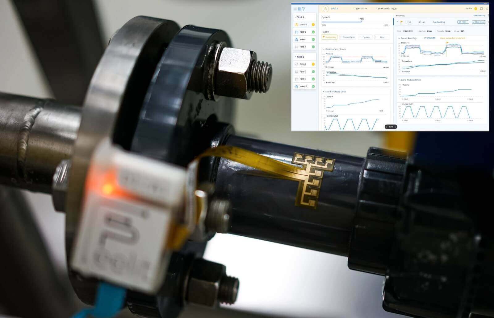
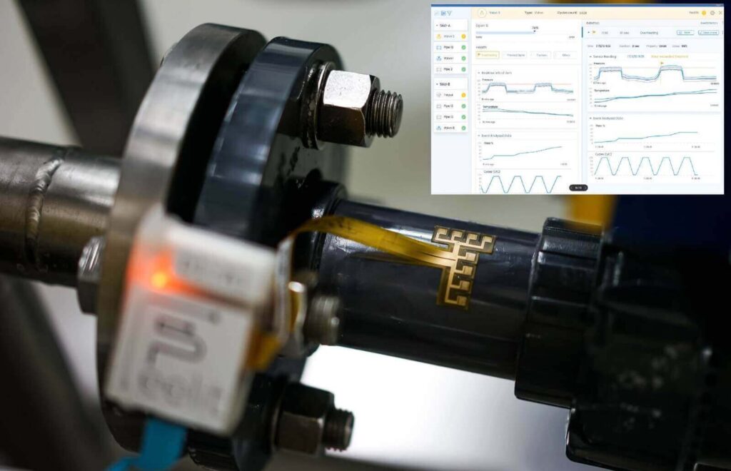
A Feelit sensor strip monitors the health of industrial equipment. Photo courtesy of Feelit
Being a vegetarian looks like its about to get a whole lot tastier. Israeli foodtech startup, Aleph Farms, which develops animal-cruelty free cultured meat products, has secured a massive $105 million Series B round. The investment was led by L Catterton, the largest global consumer-focused private equity firm, and DisruptAD. The two were joined by Skyviews Life Science, as well as a consortium of leading global food and meat companies including Thai Union, BRF, and CJ CheilJedang. Additionally, existing investors, including VisVires New Protein, Strauss Group, Cargill, Peregrine Ventures, and CPT Capital.
Juicy cruelty-free steaks
Aleph Farms has developed a system for growing beef steaks from non-genetically engineered cells isolated from a living cow, without harming animals and with a significantly reduced impact to the environment. Unlike similar products, the company’s tech doesn’t rely on genetic engineering, but rather is based on a natural process of muscle tissue regrowth in bovine. The Israeli startup has found a way to isolate the cells responsible for this process, culturing them outside of the typical host, therefore creating optimal conditions to grow beef including fat, muscle, and other features that create a juicy steak.
Considered a rare investment in the foodtech sector, Aleph’s mega-round is further highlighted due to the fact that the company’s previous funding was back in 2019 and tallied only $11.65 million, compared to the current nine-digit round. Aleph Farms states that the capital will be used to execute its plans for large-scale global commercialization of cultivated beef steaks and portfolio expansion. Additionally, a more near-term view shows the company plans on scaling-up manufacturing, growing operations internationally, and expanding its product lines and technology platform ahead of its initial market launch in 2022.
Aleph Farms’ leadership team. From left to right: Technion Professor Shulamit Levenberg, Co-Founder & Chief Scientific Adviser; Didier Toubia, Co-Founder & CEO; Dr. Neta Lavon, CTO & Vice President of R&D. Credit: Rami Shalosh (PRNewsfoto/Aleph Farms)
“This additional capital from top-tier partners with unparalleled experience and expertise brings us significantly closer to our vision of providing secure and unconditional access to high-quality nutrition to anyone, anytime, anywhere. We see our investors as partners for building this new category of meat and it was critical to us that they share our strong commitment to improving the sustainability of our global food systems,” said Didier Toubia, Co-Founder and CEO of Aleph Farms.
Aleph Farms was founded in 2017 by Didier Toubia and Professor Shulamit Levenberg. The company has raised $118 million to date. The company is definitely doing some interesting things with cultivated meat to bring more cruelty-free and sustainable options to the world, and outside of it. During October of last year, Aleph Farms launched new food driven initiative to produce cultured meat and protein solutions for Earth’s future travelers to Mars. Yep, you heard it right.
Aleph Farms completes $105 million Series B round
The company anticipates an initial market launch in 2022 and intends to scale up manufacturing, grow its global operations, and expand its product lines and technology ahead of that
Aleph Farms, an Israeli cultivated meat company that grows steaks directly from non-genetically modified animal cells, has announced the completion of a $105 million Series B funding round. The round was led by the Growth Fund of L Catterton, a consumer-focused private equity firm, and DisruptAD, one of the largest venture platforms in the Middle East. To date, it brings its total funding to $118 million.
“We are thrilled to grow our relationships with existing partners, and welcome select new investors in this funding round,” said Didier Toubia, Co-Founder and CEO of Aleph Farms. “This additional capital from top-tier partners with unparalleled experience and expertise brings us significantly closer to our vision of providing secure and unconditional access to high-quality nutrition to anyone, anytime, anywhere. We see our investors as partners for building this new category of meat and it was critical to us that they share our strong commitment to improving the sustainability of our global food systems.”
The round saw participation from Skyviews Life Science, as well as food and meat companies including Thai Union, BRF, and CJ CheilJedang. Existing investors, including VisVires New Protein, Strauss Group, Cargill, Peregrine Ventures, and CPT Capital, also took part in the round.
Aleph Farms intends to use the funds to expand its efforts for large-scale global commercialization of its cultivated beef steaks. The company anticipates an initial market launch in 2022 and intends to scale up manufacturing, grow its global operations, and expand its product lines and technology ahead of that.
Earlier this year, CTech visited Aleph Farms to explore its laboratory in Rehovot that samples cow cells, stores them in a cell bank, develops different muscle, blood vessels, and fat cells in a cell bioreactor, and then grows the tissue in a tissue bioreactor to produce a steak. “With cultivated meat, you can control the quality and tailor the quality of the raw material you get to make exactly the type of dish, cooking, and experience you want for your consumers,” Toubia told CTech at the time.
“With cultivated whole-muscle cut steaks, an optimized platform for cost parity at scale, and a global partnership network with the world’s largest meat producers, Aleph Farms has differentiated itself as the leading cultivated meat company poised to go to market,” said Michael Farello, a Managing Partner at L Catterton’s Growth Fund. “We are excited to support their success as they prepare for global launch, and we look forward to leveraging our significant expertise in food and sustainable businesses that meet the needs of a changing consumer and a changing world.”
Mansour AlMulla welcomed Aleph Farms on behalf of DisruptAD as its first Israel-based partner, and said: “Our partnership with Aleph Farms underpins our long-term desire to accelerate the path for technology pioneers and change leaders that are building technologies of tomorrow.” Mayank Singhal, also speaking on behalf of DisruptAD, added “our belief is that the future of food will be built around evolved consumer choices and a redressal of climate concerns, with Aleph playing a central role in shaping this agenda across global markets. We are delighted to partner with them.”
Aleph Farms was co-founded in 2017 by Didier Toubia, The Kitchen Hub of the Strauss Group, and Professor Shulamit Levenberg from the Biomedical Engineering Faculty at the Technion – Israel Institute of Technology. It is represented by advs. Yoav Etzyon and Efrat Shpizaizen from Amit, Pollak, Matalon & Co (APM). L Catterton has $30 billion of equity capital across its fund strategies and has made 250 investments since 1989. DisruptAD is ADQ’s venture capital platform and invests across the UAE as well as other global markets including the Middle East and North Africa region, India, China, South East Asia, and the United States.
Israel’s Aleph Farms Raises Whopping $105 Million For Cultured, Slaughter-Free Meat
Israeli cultivated meat startup Aleph Farms has raised $105 million in a Series B funding round to bring its cultured, slaughter-free meat to market next year. The company announced on Wednesday that the investment was led by the Growth Fund of L Catterton, an American-French global consumer-focused private equity firm, and DisruptAD, the venture capital arm of Abu Dhabi’s ADQ company and one of the largest venture platforms in the Middle East.
Israeli cultivated meat startup Aleph Farms has raised $105 million in a Series B funding round to bring its cultured, slaughter-free meat to market next year. The company announced on Wednesday that the investment was led by the Growth Fund of L Catterton, an American-French global consumer-focused private equity firm, and DisruptAD, the venture capital arm of Abu Dhabi’s ADQ company and one of the largest venture platforms in the Middle East.
The funding also saw participation from Skyviews Life Science, as well as a consortium of leading global food and meat companies including Bangkok-based seafood producer Thai Union, Brazilian food multinational BRF, and South Korean food company CJ CheilJedang. Additionally, existing investors, including VisVires New Protein, Strauss Group, Cargill, Peregrine Ventures, and CPT Capital, took part in the Series B funding round. The funding brings Aleph Farms’ total capital raised to date to $118 million.
Aleph Farms was founded in 2017 by Dr. Didier Toubia and Professor Shulamit Levenberg of the Biomedical Engineering Faculty at the Technion – Israel Institute of Technology, alongside Israeli food-tech incubator The Kitchen, a part of the Strauss Group.
Aleph Farms. Courtesy
In 2018, Aleph Farms unveiled the world’s first slaughter-free steak made from cow cells and a cultivated rib-eye steak earlier this year. In January, the company announced an agreement with Japanese multinational Mitsubishi Corporation’s Food Industry Group to bring cultivated meat to Japan, followed by a deal to operate in Brazil.
As a strategic partner to DisruptAD, Aleph Farms says it will evaluate the establishment of a manufacturing facility in Abu Dhabi to supply its cultivated meat products across the UAE and the broader GCC region.
Aleph Farms’ method to produce cultivated beef steaks relies on mimicking a natural process of muscle-tissue regeneration occurring inside the cow’s body, but under controlled conditions. The startup says it implements a combination of six unique technologies that allow it to drop the production costs of the meat, including innovative approaches to an animal-free growth medium to nourish the cells, and bioreactors – the tanks in which the tissue grows.
The company has also set its sights on the stars – literally – with plans announced last year for a new program called Aleph Zero. The program’s mission is to advance food security by producing fresh quality meat anywhere, independent of climate change and natural resources, and introduce new capabilities for producing food even in the harshest, most remote environments like space. This project was preceded by an experiment in 2019 to produce slaughter-free steak at the International Space Station, in a bid to demonstrate its mission to provide sustainable food security on Earth and beyond.
A rendering of Aleph Farms’ BioFarm. Courtesy
Taking Aleph Farms forward
The founders said in an announcement published on Wednesday that the fresh funding will help them execute plans for large-scale global commercialization and portfolio expansion into new types of animal protein sometime next year. The company is currently working with regulatory agencies for market entry.
“We are preparing towards an initial market launch in 2022 of our first product – a thin-cut beef steak, and are also building the foundation for global commercial expansion, including establishing long-term commercial agreements with local stakeholders from the meat sector. We have carefully hand-picked our partners based on synergies in sustainability commitments and our core values,” Toubia tells NoCamels.
Regarding the scale of the investment, Toubia says he feels “very humbled and am grateful for our partners.”
“We are thrilled to grow our relationships with existing partners, and welcome select new investors in this funding round,” Toubia, who serves as CEO of Aleph Farms, said in the announcement. “This additional capital from top-tier partners with unparalleled experience and expertise brings us significantly closer to our vision of providing secure and unconditional access to high-quality nutrition to anyone, anytime, anywhere. We see our investors as partners for building this new category of meat and it was critical to us that they share our strong commitment to improving the sustainability of our global food systems.”
The company’s near-term milestones include scaling-up manufacturing, growing operations internationally, and expanding product lines ahead of an initial market launch in 2022.
Further, says Toubia, Aleph Farms “will expand product lines and our technology platform with new animal proteins in addition to beef, and scale our 3D bioprinting platform, following the unveiling of our proof-of-concept ribeye steak in February 2021.”
“These developments are in line with our mission to generate more range and deliver a more diverse spectrum of culinary experiences associated with eating meat. We believe that additional meat designs will drive a larger impact in the mid to long term,” he tells NoCamels.
Aleph Farms’ cultivated steak. Photo: Aleph Farms
“With cultivated whole-muscle cut steaks, an optimized platform for cost parity at scale, and a global partnership network with the world’s largest meat producers, Aleph Farms has differentiated itself as the leading cultivated meat company poised to go to market,” said Michael Farello, a managing partner at L Catterton’s Growth Fund, which manages $30 billion of equity capital across its fund strategies and 17 offices around the world.
“We are excited to support their success as they prepare for global launch, and we look forward to leveraging our significant expertise in food and sustainable businesses that meet the needs of a changing consumer and a changing world,” added Farello.
Mansour AlMulla welcomed Aleph Farms on behalf of DisruptAD as its first Israel-based partner, and said: “Our partnership with Aleph Farms underpins our long-term desire to accelerate the path for technology pioneers and change leaders that are building technologies of tomorrow.”
Israel – a clean meat hub
Israel is home to a host of startups and companies working in the clean meat and/or plant-based “meat” space.
These include Future Meat Technologies, a cultured meat developer that recently launched the world’s first industrial cultured meat facility in Rehovot; Redefine Meat, an Israeli food tech startup that is carving out an important spot in the vegan meat space and recently announced a $29 million funding round that will go toward a commercial launch; SuperMeat, a developer of lab-grown poultry extracted from chicken cells; MeaTech, a cultured meat bioprinting company focused on chicken fat; and SavorEat, a vegan burger maker and the first food tech company to go public on the Tel Aviv Stock Exchange (TASE).
Aleph Farms gets $105 million investment to bring lab-grown steaks to market
Funding round is led by private equity firm L Catterton and DisruptAD, the VC arm of Abu Dhabi’s holding company ADQ; Aleph mulls manufacturing plant in UAE
Aleph Farms ribeye steak (Aleph Farms and the Technion-Israel Institute of Technology)
Aleph Farms, a maker of cultivated meat that grows steaks from modified cattle cells, said Wednesday it has raised $105 million in a Series B funding round from investors.
The round was led by the growth fund of L Catterton, a US-French consumer-focused private equity firm with over $30 billion in equity capital, and DisruptAD, the venture capital arm of the Abu Dhabi holding company ADQ.
The round also saw participation from Skyviews Life Science as well as a consortium of global food and meat companies including Thai Union, BRF, and CJ CheilJedang. Existing investors VisVires New Protein, Strauss Group, Cargill, Peregrine Ventures, and CPT Capital also participated in the latest round, bringing the funds raised by the startup to date to more than $118 million, the company said in a statement.
Aleph will use the funds to advance the global commercialization of its cultivated beef steaks and expand its portfolio of products ahead of its planned market launch in 2022, the company said.
To produce its meat, Aleph leverages the ability of animals to grow tissue muscle constantly and isolates the cells responsible. It then reproduces the optimal conditions for these cells to grow into tissue, basically growing meat outside the animal.
The tissue is grown in tanks that act as fermenters, similar to those in a brewery. There the cells are nurtured and shaped into a 3D structure that makes the meat.
DisruptAD’s investment in Aleph Farms aims to help bolster Abu Dhabi’s long-term focus on food resilience, the statement said. As strategic partners, DisruptAD and Aleph Farms will mull setting up a manufacturing facility in Abu Dhabi to supply its cultivated meat products across the UAE and the broader GCC region, the statement said.
DisruptAD invests in startups and venture capital funds and sets up new incubators and accelerators to help Abu Dhabi become a destination for startups and to accelerate the development of its own innovation ecosystem. DisruptAD invests across the UAE as well as other global markets including the Middle East and North Africa , India, China, Southeast Asia and the United States. The VC fund aims to support and nurture over 1,000 startups by 2025, the statement said.
“This additional capital from top-tier partners with unparalleled experience and expertise brings us significantly closer to our vision of providing secure and unconditional access to high-quality nutrition to anyone, anytime, anywhere,” said Didier Toubia, the co-founder and CEO of Aleph Farms.
Mansour AlMulla, chief investment officer of DisruptAD, said Aleph Farms is the firm’s first Israel-based partner, and added that the partnership “underpins our long-term desire to accelerate the path for technology pioneers and change leaders that are building technologies of tomorrow.”
Aleph Farms was founded in 2017 by Toubia, The Kitchen Hub of the Strauss Group, and Prof. Shulamit Levenberg from the Biomedical Engineering Faculty at the Technion – Israel Institute of Technology.
A Feelit sensor strip monitors the health of industrial equipment. Photo courtesy of Feelit
Moving manufacturing from “far and cheap” to “local and smart” is the goal of the so-called Fourth Industrial Revolution (Industry 4.0).
Feelit, one of many Israeli startups in the $90 billion international Industry 4.0 technology market, enables industrial equipment to communicate its maintenance needs in real time.
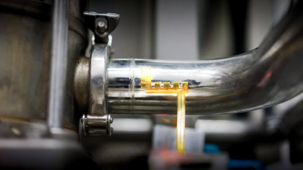
Feelit uses ultrathin flexible stickers printed with nano-ink sensors, invented by the company’s nanotech PhD cofounders.
Affixed easily to any stationary or moving object — including valves, pumps or pipes — the sensors pick up on issues such as humidity, vibration, heat, pressure or micro-fractures that could impair performance and cause costly breakdowns and interruptions.
The stickers send the collected data to the cloud for instant analysis, providing human operators with live feedback on structural and performance changes in the machinery.
If the equipment “feels” it is about to fail, it sends a request for service or replacement.
“Before our graduate studies at the Technion, we worked in the semiconductor industry so we saw how faulty valves can cause millions of dollars of damage,” says Meital Segev-Bar, referring to herself and cofounder Gady Konvalina.
“We met again in Prof. Hossam Haick’s lab at the Technion, where we worked on his electronic nose concept. Then we played around with electronic ‘skin’ that attaches to an object and makes it capable of feeling when it’s being touched and when it’s in pain, just like human skin whose job is to alert to when something is wrong.”
Easy as a sticker
These projects led Segev-Bar, Konvalina and Haick to found Feelit in 2017 in the Technion DRIVE accelerator.
They got seed funding from the Technion’s tech-transfer company and a Japanese VC, as well as the Takwin VC fund supporting northern Israeli startups.
“We see a lot of Industry 4.0 monitoring solutions for assets like motor vibration, but we found that vibration, pressure and temperature cannot be monitored continuously and noninvasively in pipe and valve systems,” says Segev-Bar, the startup’s CTO.
“We wanted a solution that could be retrofitted and installed as easily as a sticker,” she tells ISRAEL21c.
An L-shaped Feelit sticker sending data to an operator’s dashboard. Photo courtesy of Feelit
Haifa-based Feelit has finished several pilot projects with its domestically produced nano-ink stickers and is starting installations in Israel and Europe.
“It’s meant for industries that have a flow process,” Segev-Bar explains. “We specialize in assets that materials flow through.”
That means food and beverages, oil and gas, pharmaceuticals and chemicals, and any industry that uses pneumatic lines, such as auto manufacturing.
Feelit stickers also could be used in robotics and other IoT-related industries, she adds.
Investment from Henkel
The company recently concluded a $7 million Series A round in which the prominent investor is Henkel Tech Ventures of Germany.
Henkel’s adhesive materials are used in more than 800 different industries including steel mills, car factories, mining equipment and power turbines around the globe.
Feelit allows manufacturers to monitor the health of machinery in real time. Photo courtesy of Feelit
Paolo Bavaj, head of corporate venturing for Henkel Adhesive Technologies, said the company looks for novel, scalable technologies that can help traditional businesses meet increasing demands for efficient industrial IoT solutions.
“The investment in Feelit perfectly fits our business strategy and underlines the value of our long-standing engagement in Israel as a major global hub for materials and technology startups,” Bavaj said.
“With Feelit’s growth potential and Henkel’s industry expertise and market reach, our unique nanotechnology will be able to benefit a broad relevant client base,” said Feelit CEO Konvalina.
“The partnership has already begun opening up new opportunities in the oil and gas industry and working with Henkel’s MRO [maintenance, repair and operations] unit will help us to develop even more verticals and applications.”
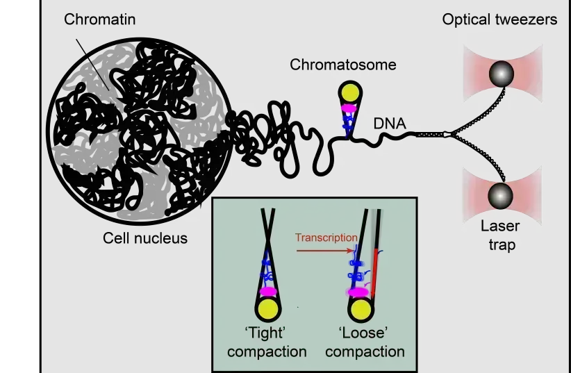
Using optical tweezer technology, Technion researchers were able to gain a greater understanding of the poorly understood DNA packaging process, which impacts how genes are expressed.
Optical tweezers apply force on DNA, and “unzip” it into two separate strands. Upon reaching the chromatosome the unzipping is halted by contacts of the histone proteins (yellow, pink, blue) with the DNA, revealing whether the chromatosome is in an “open” (right) or “closed” (left) structure.
(photo credit: TECHNION)
Scientists from the Technion-Israel Institute of Technology have used “laser tweezers” to understand the structure of DNA better than ever before, shedding light on poorly understood mechanisms that influence how genes express themselves in the human body.
Chromatin is found in DNA, the essential code in the human body that provides instructions needed for function and development. Though there is said to be around two meters total of DNA in the human body, found in the nucleus of every single cell, it is compressed into just tens of microns in size. This is because of how DNA itself is packaged, formed into a compact structure known as chromatin.
Chromatin itself is organized by wrapping the DNA strands around a specific protein called histones. The spool-like structure it eventually forms is said to resemble beads on a string. These “strings” are then connected with a special type of histone called a linker histone, which helps the strands form into more complex structures called chromatosomes.
The advantage of packaging the human genome in this form is that it makes it possible for it to actually physically fit within the cell. However, it makes accessing it more difficult, which can pose problems for mechanisms within the cell that are supposed to read the DNA.
The result is that the way a gene is ultimately expressed becomes dependent on a particular method of packaging. How exactly this works is still a mystery, and scientists have struggled to find an answer. But one thing that has been uncovered is the role of linker histones in the organization of this packaging. In other words, if a linker histone malfunctions, it could lead to improper packaging, which can result in non-ideal gene expression.
According to some experts, it is believed that the end result of linker histone malfunctions could manifest as autism or serious diseases like cancer. But how the linker histones actually bind DNA in the first place remains a mystery, making it even more difficult to investigate the issue using conventional methods.
But an unconventional method is exactly what Dr. Sergei Rudnizky used. The scientist and his team developed a new method based on “optical tweezers,” a method that uses a focused laser beam to capture individual molecules and exert force on them.
The method itself was pioneered by Jewish-American scientist Arthur Ashkin in 1986. It was based on years of research he had conducted in the 1970s, which had later formed the basis for the 1986 work by Steven Chu on using optical tweezing on cooling and trapping neural atoms. Chu would win the Nobel Prize in Physics for this in 1997, and would later go on to serve as US secretary of energy from 2009 to 2013. Ashkin himself would later win the Nobel Prize in Physics for it in 2018.
The technology was also later employed in other sectors, and in 2010 was adopted by Tel Aviv University for nanotechnology research.
With this laser tweezer, Rudnizky, under the supervision of Profs. Ariel Kaplan and Philippa Melamed were able to slowly detach one strand of DNA from the rest of the strands. The process functions in a manner similar to unzipping a zipper, slowly removing the strand from the chromatosome. It isn’t a smooth process though, as the DNA strand can get stuck if it makes even the slightest contact with a histone. When that happens, more force is applied to advance further.
And as it turns out, histone-DNA contact is far more extensive, with chromatosomes being much larger than was previously believed. The linker histones themselves were also surprisingly flexible in structure because there were two different shapes the chromatosome can shift between, a symmetric and compact one and a relaxed and asymmetric one.
But it is possible to externally control the transition between these shapes through transcription mechanisms in the cells.
As suggested in the findings, published in the academic journal Molecular Cell, it is possible that the cell utilizes this transition to regulate its access to the DNA.
These findings are extremely significant, as they shed light on poorly understood functions of genome expression, which can further knowledge on the role of chromatin and chromatosomes in health and diseases.
HAIFA RESEARCHERS USE LASER ‘TWEEZERS’ TO STUDY STRUCTURE AND DYNAMICS OF DNA PACKAGING IN ALL CELLS
Bind them as a sign on your hand and let them serve as a symbol on your forehead.
Each one of the cells in our bodies contains DNA, which dictates the instructions needed for living things to develop and function. Incredibly, a total of two meters of DNA is packaged in each tiny cell’s nucleus, which is just tens of microns in size. This is made possible to packaging the DNA into a compact structure called chromatin.
The basic level of chromatin organization is provided by wrapping the DNA around proteins called histones in a spool-like structure that resembles beads on a string. Then, more complex structures called chromatosomes are formed with the help of a special histone known as a linker histone that connects the strings.
But scientists have not known much about the structure and dynamics of chromatosomes, leaving the most basic questions of how they bind DNA.
Now, researchers in the Biology Faculty of the Technion-Israel Institute of Technology in Haifa have used “laser tweezers” to accomplish this. Their study, just published in the journal Molecular Cell under the title “Extended and dynamic linker histone-DNA Interactions control chromatosome compaction,” was conducted by Dr. Sergei Rudnizky under the supervision of Profs. Ariel Kaplan and hilippa Melamed.
Packaging of the genome is essential for it to fit into the cell, but it also reduces the accessibility to the cellular machines that read the DNA and transcribe the genes. Therefore, the distinct packaging at a particular gene will have a huge impact on its expression in ways that are only beginning to be unraveled.
In particular, linker histones are known to play a key role in this organization of the genome – and their malfunction can lead to serious diseases including cancer and autism.
The lack of understanding of these crucial processes stems from the dynamic nature of linker histones, which makes it challenging to investigate them using conventional methods based on sampling a huge number of molecules simultaneously.
Kaplan’s lab developed a unique method based on “optical tweezers” – an approach that allows researchers to capture individual chromatin molecules and exert forces on them with the help of a focused laser beam.
In these experiments, one strand of DNA is slowly detached from its complementary strand in a manner similar to a zipper being unzipped, through the entire structure of a chromatosome. The principle of the measurement is simple – at points where a histone makes contact with the DNA, even in the weakest way, the zipper gets stuck, and more force needs to be applied to overcome the histone-DNA contact and advance into the structure.
Using this approach, the team discovered that contacts between histones and DNA are far more extensive than previously known and that chromatosomes are, in fact, much larger than previously thought. They also they found a surprising flexibility in the structure of linker histones, as two different chromatosome shapes exist – one symmetric and compact and the second asymmetric and more relaxed.
Amazingly, transition between these shapes in an individual molecule can be externally controlled by the transcription machinery itself. This suggests that the cell uses the transition between stable and unstable forms of a chromatosome to regulate access to the DNA in a controlled manner. Given the key role played by chromatosomes in maintaining proper expression of our genome, these findings add an important layer to our understanding of the role of chromatin architecture in health and disease.
A new artificial intelligence system can diagnose heart issues with greater precision than people.
Article published at www.jpost.com on July 12, 2021.
3D image of a heart in a cardiology test
(photo credit: REUTERS)
Technion researchers have discovered a viable way in which to reliably use Artificial Intelligence (AI) in medicine and demonstrated the use of practical systems for cardiology in an article published in Proceedings of the National Academy of Sciences of the United States of America (PNAS).
Even as AI has developed greatly over the past decades, the use of this technology in medical products is still scarce, and the methods currently employed by doctors are based on older technology.
In the article, the research team demonstrated the use of the new technology to identify diseases based on hundreds of electrocardiograms (ECG), which are currently the most common technology used in cardio-medicine.
The new systems analyzes the ECGs using an augmented neural network, which has been taught various patterns based on a system of more than 1.5 million ECG tests from hundreds of patients in hospitals worldwide.
The ECG is a quick and non-invasive test that provides information on the workings of the heart. The disadvantage of the test is that the cardiologists who read the printouts are susceptible to mistakes in their interpretations, either because they are subjective or because they cannot see what they are looking for with enough precision.
The new systems are more accurate and can detect pathological conditions that human cardiologists are physically unable to see.
The researchers worked closely with cardiologists and created the system according to their requirements. The output includes an uncertainty estimation of results, alerts regarding inconclusive results and increased risk of pathology that the ECG signal does not observe itself.
The system demonstrated enough sensitivity and precision that it can alert for risk of arrhythmia even when it is not demonstrated in the ECG. Without this early diagnosis, people run a higher risk of heart-attacks and strokes.
Moreover, the AI uses official cardiology terminology to explain its decisions.
The study was headed by Prof. Yael Yaniv, director of the Bio-electric and Bio-energetic Systems Laboratory at the Faculty of Biomedical Engineering at the Technion; Prof. Alex Bronstein, director of the VISTA Laboratory at the Taub Faculty of Computer Science; Prof. Assaf Schuster of the Learning at Scale Laboratory (MLL) at the Taub Faculty of Computer Science and co-director of the MLIS Center (Machine Learning & Intelligent Systems); Yonatan Elul, a doctoral student in the laboratories of Professors Bronstein, Yaniv, and Schuster and Aviv Rosenberg, a doctoral student in the laboratory of Professors Bronstein and Yaniv.
The project was sponsored by the Ministry of Science and Technology and the Technion Hiroshi Fujiwara Cyber Security Research Center and the Israel Cyber Directorate.
ISRAELI RESEARCHERS TEACH COMPUTERS TO READ AND ANALYZE ELECTROCARDIOGRAMS BETTER THAN CARDIOLOGISTS
And they told him, “Yosef is still alive; yes, he is ruler over the whole land of Egypt.” His heart went numb, for he did not believe them.
Man can teach computers to do many things better than humans can today; in fact, computers can teach other computers to do so. In recent years, spectacular progress has been made in the world of artificial intelligence (AI) and “deep learning” (machine-learning methods based on artificial neural networks that have been applied to fields including computer vision, speech recognition, natural language processing, machine translation, bioinformatics, medical image analysis, material inspection and board game programs, where they have produced results comparable to and in some cases surpassing human expert performance).
Despite their great promise, AI systems have yet to become ubiquitous in the daily practice of medicine largely due to several crucial unmet needs of healthcare practitioners. These include lack of explanations in clinically meaningful terms, handling the presence of unknown medical conditions and transparency regarding the system’s limitations, both in terms of statistical performance as well as recognizing situations for which the system’s predictions are irrelevant.
So at present, there are virtually no medical products on the shelf that use this technology, and as a result, doctors continue to use the same tools used in previous decades.
Prof. Yael Yaniv (Technion)
To find a solution to this problem, the research group of Prof. Yael Yaniv of the Faculty of Biomedical Engineering at the Technion-Israel Institute of Technology in Haifa joined forces with the research groups of Prof. Alex Bronstein and Prof. Assaf Schuster of the Taub Faculty of Computer Science to create an AI-based system that automatically detects disease on the basis of hundreds of electrocardiograms, which are currently the most widespread technology employed for the diagnosis of cardiac pathology.
Elul and Aviv Rosenberg Now, under their joint supervision, research by doctoral students Yonatan has been published in Proceedings of the National Academy of Sciences of the United States of America (PNAS) under the title “Meeting the unmet needs of clinicians from AI systems showcased for cardiology with deep-learning–based ECG analysis.
The new system automatically analyzes the electrocardiograms (ECGs) using augmented neural networks – the most prominent tool in deep learning today. These networks learn different patterns by training on a large number of samples, and the system developed by the researchers was trained on more than 1.5 million ECG segments sampled from hundreds of patients in hospitals in various countries.
The electrocardiogram, developed more than 100 years ago, provides important information on conditions affecting the heart quickly and non-invasively. The problem is that the printouts have to be interpreted by a human cardiologist, so their interpretation is – by necessity – pervaded by subjective elements. As a result, numerous research groups around the world are working to develop systems that will automatically interpret the printouts efficiently and accurately. These systems are also able to identify pathological conditions that human cardiologists –
regardless of their experience – are unable to detect.
The Technion system was built according to the needs set down by cardiologists, and its output includes an uncertainty estimation of the results, indication of suspicious areas on the ECG wave and alerts regarding inconclusive results and increased risk of pathology not observed in the ECG signal itself.
The system is sensitive enough to provide alerts regarding patients at risk of arrhythmia (irregular heartbeat) – even when the arrhythmia is not shown on the ECG printout. In addition, the rate of false alarms is very minimal. The new system also explains its decisions using the accepted cardiology terminology.
The researchers hope their system can be used for cross-population scanning for the early detection of those who are at risk of arrhythmia. Without this early diagnosis, these people have an increased risk of heart attack and stroke.
Humans, unlike some animals, have a limited ability to regrow missing body parts. However, pioneering research at Technion University could change this.
Faculty of Materials Science and Engineering at Technion University.
(photo credit: Wikimedia Commons)
Technion University researchers are tackling one of the most pressing medical issues: loss of tissue.
Prof. Shulamit Levenberg and her team in the Biomedical Engineering Department have pioneered research that could make it easier for humans to cultivate tissue, Technion UK stated in a press release on Monday.
Levenberg said she is currently working on autografting to make this a reality, which is the procedure of moving tissue from one part of the body to another. However, new pioneering research from the Technion might make this process a lot easier.
This is just one of many new innovations that are coming out of the Technion, as the school has been considered the leading institution for Israeli innovation.
Long-term implications of Levenberg’s could lead to allowing patients to receive bone and tissue matter in a vat, rather than needing to remove it from another part of their body.
Alan Aziz, CEO of Technion UK, said that the process of humans growing their own body-part replacements in a lab sounds like science fiction, but may soon become a reality.
“It may soon become reality thanks to pioneering research at the Technion – and that’s the tooth, the whole tooth, and nothing but the tooth!” he said.
Just last month, the Technion came out with another groundbreaking innovation that can quickly diagnose tuberculosis.
Tissue autografting turns pulp fiction into pulp reality
One of the most challenging medical conditions is serious loss of tissue. Unlike certain animals, humans have only a limited capacity for regrowing missing body parts, which is how the science of auto-grafting has been developed. Moving tissue from one part of the body to another is still complicated though – but pioneering research from Israel may be about to make it easier.
Autografting revolves around replacing a missing piece of the body with another. For example, if there has been a significant loss of bone in a hip or knee which means that section of the skeleton can no longer repair itself, a piece of rib could be grafted in instead. The key to success is the supporting infrastructure of soft tissue and blood vessels that enables the bone to heal. This material therefore also needs to be transplanted from one area of the body to the other.
This is the issue that Professor Shulamit Levenberg is taking on. Levenberg works in the Faculty of Biomedical Engineering at the Technion – the Israel Institute of Technology. Along with her colleagues, she is part of a lab that is looking to grow this specialised tissue in vats so that it can be used for auto-grafting. This lab has now successfully built soft tissue with blood vessels from dental pulp, which is the material inside teeth. The stem cells from the dental pulp help blood vessels to form, supporting tissue growth and healing.
This new procedure has been trialled in repairing a bone defect in rats, inserting specially cultivated tissue rather than the traditional method of using existing tissue from another part of the body. This new tissue is deemed to have been more effective in enabling the surgery to heal, whilst also not needing to create another wound in order to extract material to be transplanted, as is usually the case.
In the future, this research could allow patients to receive bespoke bone and tissue matter created in vats, rather than needing to remove it from another part of their body. Where this to be case, it would represent another significant contribution to medical science by the Technion. Since 1912, the academic institution has been at the forefront of spearheading Israel’s scientific endeavours. Israel today is the country with the highest percentage of scientists and engineers – and the majority of them studied at the Technion, home to three of Israel’s five science Nobel Laureates.