A hundred million patients the world over benefit from the technology of Diagnostic Robotics — most of them are not even aware of it. Via artificial intelligence and big data analysis, the company from Israel is trying to make the world’s health systems more efficient and to predict the patients’ deterioration. “There are very few opportunities in life to do something with such a big impact.”
Article published at www.forbes.co.il on June 29, 2021.

Total fundraising: $38 million
Employees: 110 employees; 80 of them in Israel
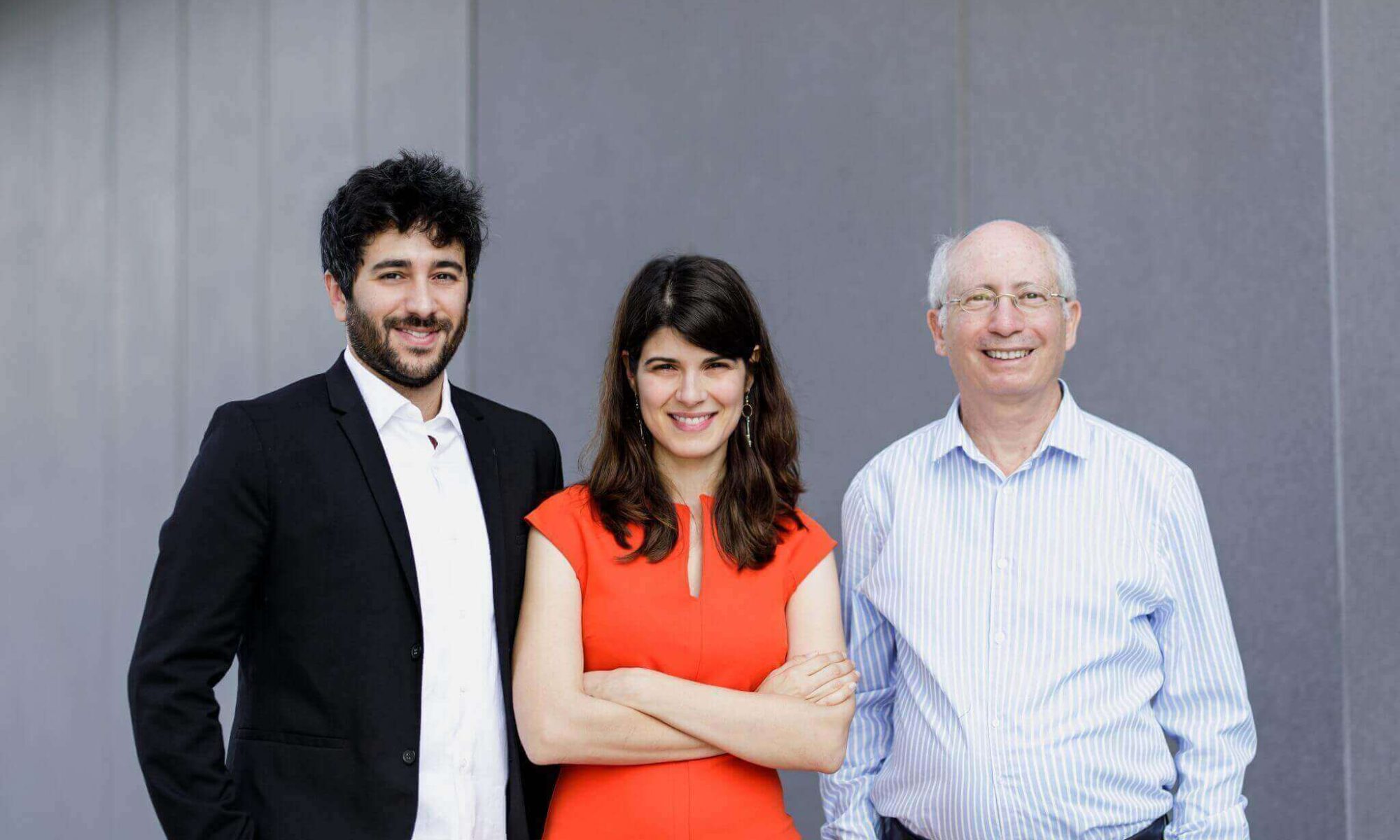
Founders: Yonatan Amir, Kira Radinsky And Moshe Shoham
Founded: 2017
Investors: Mayo Clinic, Maverick, Accelmed, Kobi Richter, Morris Kahn and others
“Throughout the pandemic we all saw what happened when there was a heavy load placed on the health system. In another ten years this load will naturally accumulate and reach similar dimensions to those that we saw during COVID-19,” predicts Dr. Kira Radinsky, acclaimed scientist and alumna of the Forbes 30 Under 30 list.
Radinsky (34), got her start in the world of artificial intelligence and big data at the young age of 15, when she became a student at the Technion. During her doctorate she developed mechanisms for predicting events based on gathering information from news sites and social networks, and among other things, predicted the outbreak of the cholera epidemic in Cuba in 2012. Since then she has added more accomplishments to the list, including selling the start-up SalesPredict, where she served as VP of technology, to eBay.
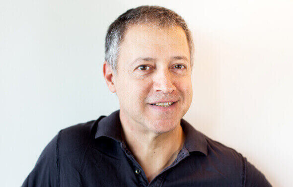
In 2017 she met with Yonatan Amir (33), also an alum of Forbes Israel’s 30 under 30 list, and Professor Moshe Shoham (founder of Mazor Robotics) and together they founded Diagnostic Robotics — a start-up based on AI technology which assists medical teams to make faster and more accurate diagnostic decisions, and to provide physicians with vital data that will help them make the best decisions. For example, a doctor who receives a patient at the hospital can be assisted by the system in order to quickly determine a triage score for the patient (the index which determines the urgency of treatment).
With the coronavirus outbreak in Israel, another initiative took shape — using Diagnostic’s technology to identify hotspots. In cooperation with the Ministry of Health, the HMOs and other research bodies, a symptoms questionnaire was distributed to the public which helped decision-makers to process data and curb outbreaks in various parts of the country.
“We are looking at the coronavirus system as a triage system in every way. The symptoms which were knowns then to be coronavirus were fever and cough, and by analyzing the answers through our system, within three days we also identified the symptom of loss of taste and smell,” says Radinsky, company chairman. “It was among the insights that we brought to the HMOs and it really changed the triage process because there weren’t enough tests, and in general the health system had to know how to prioritize the division of resources.”
In general, the coronavirus was a boon for the company, as the field of digitalized medicine received a huge boost. “There is a very large wave of inquiries from organizations from the fields of digital medicine, remote consulting and early forecasting,” says Amir, CEO. “With the first wave there was a huge drop in the number of emergency room visits — 42% less. If the medical problem wasn’t very severe, people wouldn’t come… suddenly there was a spike in the use of digital medical systems, alongside the natural development of consumption habits.”
What are the chances?
Diagnostic’s core business is roughly divided into two parts: on the patient side, the company operates technology for regulating loads through automation in the reception of patients’ data, rapid diagnosis and referrals for further treatment in health institutions; on the health organizations’ side, the company developed a system for preventative medicine, based on the organizations’ databases and the broader historical picture — thanks to which it provides systems with early detection of patients’ chronic diseases, and provides warnings to health organizations. “For example, if a diabetic patient is not treated in time, he will deteriorate to the point that he will turn up at the emergency room,” Amir offers by way of example and clarifies that the price of such a development is paid by both the patient and the institution administering treatment.
“We work across the whole spectrum. In working with health organizations, there are those for whom we are the first to help them with the processes of digitization and automatization, and there are organizations who take our product as a kind of ‘plug and play.’ It’s like a puzzle that can be put together or taken apart.”
“We are building the capacities for medical decision-making so that it can be used to establish an order of actions,” Radinsky continues. “Let’s say I have pain in my lower belly now, and my day is such that I have Zoom calls all day. Just to make an appointment, to get to the doctor and wait for my turn — it’s a whole production. We think about how it could be possible to save ourselves some of the appointments. While booking a doctor’s appointment, the application can ask me. Behind the scenes we can offer a doctor the probability, what percentage it is likely that I have a urinary tract infection. And the HMO decides at what probability a urine test referral should be done, and what probability the doctor should look at the results within three hours and approve antibiotics remotely. So what did we do? We didn’t build the application for the HMO, we didn’t build the digital system for the HMOs, but our tools are in there.”

Diagnostic’s systems integrate a variety of sources of data in real time: from sensors on the body of the patient, to lab test results to analysis of his medical records in the computerized medical file. Naturally, this is a learning system which is constantly improving, and it is important for the company’s people to emphasize that this is anonymous information coming from different health organizations. Israel, by the way, has the second largest medical database in the world.
“In recent years we received access to several billion past medical visits, and we developed tools that know what to ask the patient, to predict how urgent the care is and which tests he needs. We made it possible to automize the medical system. A family doctor today is responsible for over 2,000 patients, and he needs to be responsible for them in a proactive way too: to see deterioration and predict it in time, but that is very hard to do in an efficient way when you have thousands of patients,” says Radinsky.
“We are a company of deep-tech, automation and early detection. What is important to us is to be the necessary piece of the puzzle for leading health organizations. During the pandemic, we connected with additional corporations and sold our technology to governments. We developed different kinds of systems and we want to implement them on top of an existing set of services of health organizations and improve patients’ quality of life,” says Amir.
Touching millions
Among the company’s clients are hospitals, insurance companies, governments and commercial companies in Israel and around the world, including: Mayo Clinic, Salesforce, Deloitte, the Israeli Ministry of Health, Clalit Health Services [HMO], Leumit Health Services {HMO] and others.
The main market that the company is targeting is in the US, where healthcare spending crossed the 3.8 trillion dollar mark last year: Americans spend $460 to 680 billion dollars a year on emergency medical services, and an average of $350 billion annually on family medicine.
Within about three years, the analysts predict, the general spending on health services will grow to $5.5 trillion. If patients with fitting symptomatic and diagnostic segmentation are referred for a visit to the family doctor as opposed to a specialist — the annual savings are expected to be $170-250 million.
“100 million patients benefit from our technology, through dozens of health organizations all over the world. The company’s sales began officially in 2019, after two years of building the product, and everything grew very rapidly, with the main pricing model being SaaS (Software as a Service).”
110 workers are employed by Diagnostic Robotics: 80 of them are in Tel Aviv, in research and development roles, and 30 in New York are responsible for the marketing, sales and business development. Up until now, funding of $38 million was raised in a series A round, a big part of which was in November 2019, after which the company raised 24 million dollars led by Accelmed , Maverick, Mayo Clinic, billionaires Kobi Richter and Morris Kahn and others.

“By the end of the year we want to have a major growth round led by leading funds and strategic investors,” says Amir. “Furthermore, we want to continue to grow the rate of revenue and the number of customers, and to reach more organizations, particularly in the American market. We already reached the cream of the crop of the US businesses, so now our goal is how to begin to work in a top-down approach, and to begin to supply smaller organizations and deploy systems at a rapid pace.”
“We are about to publish new scientific articles. One of them shows how our technology successfully anticipates medical deterioration as much as a half year or a full year in advance, and this has great clinical value,” Radinsky adds. “We are talking about a clinical trial on a very large scale — in other words, it gives it the clinical seal of approval. For the first time we have data from billions of past visits and can save the lives of many, and if not save them then at least extend many lives by at least five years.”
According to Radinsky, the success, as they see it, is in the fact that they are touching hundreds of millions of people’s lives, and not the value of the company. “The clinical impact and also the business growth of the company is what is important — not the rounds of funding.”
Amir displays a more business-oriented approach. “In the end, in order to build big companies, the company really needs a strong financial backbone, the kind that can fund more and more innovation and products. We are in territory where we can built a company of a few billion dollars. If at another time it was unusual to speak of unicorns — today it happens at a high speed, for a lot of reasons. We know how to get there in a relatively simple and clear way. We are strong technologically, and are at the forefront of companies providing advanced technology.”
“I think that there are very few opportunities in life to do something with a big impact that will totally change the worldview in such an important field — like the one in which we work. I am proud that we are in this field, the impact that we are making, and that the company is also growing significantly on the business level in order to help us make more and more of an impact,” Radinsky concludes.


 Faculty of Materials Science and Engineering at Technion University. (photo credit: Wikimedia Commons)
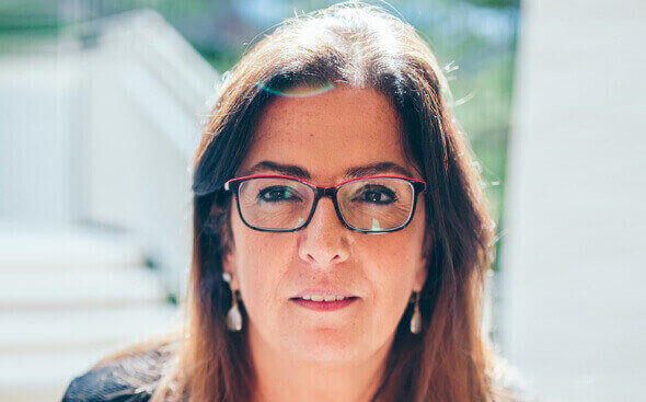
 NanoGhost co-founder and CEO, Yonatan Malca Photo: Courtesy

 Ford autonomous vehicle. Photo by Ruslan Berdichevsky.
 The SAIPS team. Photo by Dana Berman.
 Rotem Littman, co-founder of SAIPS
 Ford Motor Company Executive Chairman Bill Ford opens the Ford Research Center in Tel Aviv. Photo by Simona Shemer.