Unlike artificial language models, which process long texts as a whole, the human brain creates a “summary” while reading, helping it understand what comes next.

In recent years, large language models (LLMs) like ChatGPT and Bard have revolutionized AI-driven text processing, enabling machines to generate text, translate languages, and analyze sentiment. These models are inspired by the human brain, but key differences remain.

A new Technion-Israel Institute of Technology study, published in Nature Communications, explores these differences by examining how the brain processes spoken texts. The research, led by Prof. Roi Reichart and Dr. Refael Tikochinski from the Faculty of Data and Decision Sciences. It was conducted as part of Dr. Tikochinski’s Ph.D., co-supervised by Prof. Reichart at Technion and Prof. Uri Hasson at Princeton University.

The study analyzed fMRI brain scans of 219 participants while they listened to stories. Researchers compared the brain’s activity to predictions made by existing LLMs. They found AI models accurately predicted brain activity for short texts (a few dozen words). However, for longer texts, AI models failed to predict brain activity accurately.

The reason? While both the human brain and LLMs process short texts in parallel (analyzing all words at once), the brain switches strategies for longer texts. Since the brain cannot process all words simultaneously, it stores a contextual summary—a kind of “knowledge reservoir”—which it uses to interpret upcoming words.

In contrast, AI models process all previously heard text at once, so they do not require this summarization mechanism. This fundamental difference explains why AI struggles to predict human brain activity when listening to long texts.

To test their theory, the researchers developed an improved AI model that mimics the brain’s summarization process. Instead of processing the entire text at once, the model created dynamic summaries and used them to interpret future text. This significantly improved AI predictions of brain activity, supporting the idea that the human brain is constantly summarizing past information to make sense of new input.

This ability allows us to process vast amounts of information over time, whether in a lecture, a book, or a podcast. Further analysis mapped brain regions involved in both short-term and long-term text processing, highlighting the brain areas responsible for context accumulation, which enables us to understand ongoing narratives.

Prof. Shai Shen-Orr of the Ruth and Bruce Rappaport Faculty of Medicine showcased his lab’s trailblazing efforts in harnessing computational tools and innovative methodologies to redefine our understanding of the immune system. A leading immunologist and director of the Zimin Institute of AI Solutions in Healthcare at the Technion and the new Technion Institute for Healthy Aging, his work spans from developing metrics like “immune age” to spearheading global health projects, promising transformative implications for medicine.

Immune Age and Predictive Medicine

One of Prof. Shen-Orr’s standout contributions is the concept of immune age, a metric that quantifies the immune system’s state. This marker has shown predictive power for various health outcomes, including cardiovascular disease, paving the way for early diagnosis and intervention.

“Your immune system is a learning system,” Shen-Orr explained, emphasizing how the immune system evolves over time and adapts to environmental challenges. “By understanding an individual’s immune age, health care providers can better predict and manage potential health issues, leading to more personalized and effective treatments.”

Developed using advanced mass cytometry and machine learning, this metric represents a leap in precision medicine, shifting the focus from general health indicators to immune-specific markers.

Bridging the Data-Insight Gap With AI

Prof. Shen-Orr’s research tackles a critical bottleneck in biomedical science: the gap between vast amounts of data and actionable insights. He has pioneered computational disease models that leverage artificial intelligence to improve drug development efficiency. For example, his lab’s algorithm, “Found in Translation,” enhances the predictability of findings from animal models, like mice, to human systems by up to 50%. “We train the computer system to learn the difference between a mouse and a human,” he said.

“Most drugs fail during development. It costs about $2.5 billion to bring a single drug to market, primarily because of failed trials. By improving the translational accuracy between species, we can reduce time, cost, and animal use significantly.”

The Human Immunome Project: A Global Collaboration

One of his most ambitious initiatives, Shen-Orr is co-chief science officer of the Human Immunome Project. This global nonprofit aims to map baseline immune variations across populations, genders, and geographic regions, addressing a fundamental gap in immunological research. The project holds the potential to revolutionize vaccine development and personalized immunotherapies by understanding how different immune systems respond to treatments.

An example that underscores this need is the malaria vaccine. While it demonstrated over 90% efficacy in trials conducted in the U.S., its effectiveness dropped to less than 20% in African populations, a disparity attributed to baseline immune variations.

“We’re at a singularity moment in immunology,” Shen-Orr said. “The tools to measure the immune system comprehensively, along with AI capabilities, have matured. Now is the time to leverage them for global impact.”

From Academia to Real-World Applications

The Technion’s emphasis on translational research is evident in Shen-Orr’s dual roles as an academic leader and entrepreneur. He co-founded CytoReason, a company that integrates AI-driven disease models into pharmaceutical research and development. The platform has already gained traction with leading industry players like Pfizer and Sanofi, demonstrating its potential to streamline drug development and reduce costs.

Moreover, his collaboration with other Technion researchers is pushing the boundaries of innovation. For instance, partnerships exploring the impact of diet on immune health are underway, aiming to create tailored nutritional solutions for aging populations.

Shen-Orr also advocates for equipping biologists and clinicians with computational and data science skills to harness the explosion of data in immunology. The Technion’s curriculum now incorporates quantitative thinking from the first year of medical school, preparing future physicians to engage with cutting-edge technologies.

Looking Ahead

Prof. Shen-Orr’s work exemplifies the Technion’s commitment to groundbreaking science with tangible societal benefits. By bridging biology, AI, and global collaboration, his research not only advances our understanding of the immune system but also lays the foundation for a future where medicine is predictive, personalized, and precise.

His collaborative efforts with institutions like Stanford University and the National Institutes of Health aim to deepen our understanding of immune health and develop new diagnostic tools and therapies. “We are studying the effects of the environment and pollution on immune health,” for example.

As he puts it, “We’re moving toward a world with less trial and error and more informed decisions in medicine. The immune system, with all its complexity, is the key to unlocking this future.”

From predictive medicine to global health initiatives, Shen-Orr’s work is paving the way for a deeper understanding of the immune system and its applications in improving human health. As the field continues to evolve, the Technion remains at the cutting edge, driving innovation and collaboration in immunology research.

Proteins, the pillars of cellular function, often assemble into “complexes” to fulfill their functions. A study by the University of Geneva (UNIGE) and the Weizmann Institute, in collaboration with the Technion, reveals why this assembly often begins during the very process of protein synthesis or “birth.”

These early interactions involve proteins whose stability depends on their association. They can be compared to a couple in which each partner supports the other. This model paves the way for new strategies to understand and correct assembly errors, which are often associated with pathologies, including neurodegenerative disorders and certain cancers. The findings are published in the journal Cell.

Proteins are large molecules composed of a chain of amino acids. They are produced by the ribosome, a cellular “machine” that reads the instructions contained in messenger RNAs. Once the protein is formed, interactions between the amino acids induce the chain to fold onto itself and adopt a specific structure. While some proteins function independently, many must assemble with specific partners into complexes to fulfill their roles.

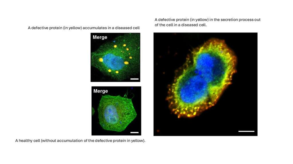
The formation of these complexes is a delicate process. If proteins fail to find their partners or fold incorrectly, this can lead to cellular dysfunction and pathologies such as Alzheimer’s disease or certain cancers. Until very recently, scientists believed that proteins only formed complexes after being fully synthesized (post-translational assembly).

However, the recent study revealed that assembly between nascent proteins—co-translational assembly—is widespread. This study identified thousands of proteins involved but did not determine the specific pairs of proteins formed or the molecular signatures underlying this early recognition.

Thousands of protein structures analysed

The group led by Emmanuel Levy, a full professor in the Department of Molecular and Cellular Biology at the UNIGE Faculty of Science—previously a professor at the Weizmann Institute—in collaboration with the group of Ayala Shiber, a professor at the Technion, focuses on the fundamental principles governing protein self-organization. In other words, these scientists aim to identify the general rules of protein assembly.

For this study, the team analyzed a list of proteins involved in co-translational assembly. By comparing their structures to those of proteins that assemble after translation, they were able to establish fundamental differences between these two mechanisms

“Our bioinformatics analyses revealed that proteins interacting with their partners while still being synthesized tend to be unstable when isolated. These proteins depend on their partners and if they do not find it, they adopt a wrong shape and get degraded,” explains Saurav Mallik, a researcher at the Weizmann Institute and co-first author of the study.

A predictive model

“Using this approach, we developed a model based on a large corpus of structural data, using both experimentally determined structures and those predicted by the artificial intelligence software AlphaFold. Our model leveraged structural properties of a complex to predict whether it associated co- or post-translationally,” add Johannes Venezian and Arseniy Lobov, co-first authors of the study. The scientists notably discovered that binding sites are exposed early in these proteins, enabling them to interact with their partner shortly after emerging from the ribosome.

These predictions were validated using experimental data focused on several proteins. “These findings pave the way for a better understanding of protein assembly within cells and highlight the global impact of protein structure on the regulation of their synthesis,” says Levy.

Many diseases, including neurodegenerative disorders and certain cancers, are linked to misfolded proteins or defective complexes. By understanding the rules of co-translational assembly, scientists could develop strategies to prevent these errors and design new therapeutic approaches to correct them.

What if we could provide groundbreaking accessibility solutions to people with disabilities?

How can technology be harnessed to offer unique solutions to people with disabilities? The Technion has designed a new course to empower students to do just that through social-technological entrepreneurship. Open to all Technion students as well as University of Haifa physiotherapy students, the course fosters interdisciplinary collaboration to address real-world challenges.

Students will explore topics such as accessibility, the psychology of people with disabilities, and principles from biomedical engineering, physiotherapy, and occupational therapy. Visits to Loewenstein Rehabilitation Hospital and Sheba Medical Center will provide firsthand insights into rehabilitation needs, enriching the learning experience.

Dr. Yacov Malinovich, the course leader, highlighted its timely significance: “Awareness of the needs of disabled people has increased, and this has become even more important in light of the ongoing war. Developing suitable technologies for rehabilitation offers students and engineers an opportunity to directly improve people’s well-being. Israel has extensive knowledge in this field, with examples like unique wheelchairs, emergency bracelets, and special surfboards.”

Dr. Malinovich, a founder of Haifa3D, brings expertise in designing assistive devices for individuals with upper limb disabilities. Haifa3D’s impactful work includes creating robotic hands for children and collaborating with the Technion’s Biorobotics and Biomechanics Lab to develop customized solutions.

“The new course will feature guests from various academic and rehabilitation institutions,” explained Dr. Malinovich. “By connecting with rehabilitation centers and individuals with disabilities, we aim to create technological solutions that truly assist those in need. Each student team will submit a product as their final project.”

Held in the Faculty of Mechanical Engineering, the course is a collaboration between t:hub – the Technion Innovation and Entrepreneurship Hub, the University of Haifa’s Physiotherapy Department, and the Technion Social Incubator. Offering six academic credits, it provides students with hands-on experience to develop innovative solutions that can transform lives.

“Stay positive,” we’re told when suffering from an illness. It’s easy to dismiss such comments as platitudes from well-meaning friends. But Technion scientists have demonstrated that activation of the brain’s reward system can boost recovery from a heart attack. Establishing the connection between the two can potentially lead to therapeutic avenues for intervention.

“It’s time that both researchers and clinicians take the link between psychology and physiology seriously,” said Technion Associate Professor Asya Rolls, a psychoneuroimmunologist and pioneer in mind-body interactions.

Scientists have previously shown that the emotional state can influence the course of disease following a heart attack. But until now, the underlying physiological mechanisms were not well understood.

Prof. Rolls worked with renowned cardiac researcher Professor Lior Gepstein and Hedva Haykin, Ph.D. ’23, in the Ruth and Bruce Rappaport Faculty of Medicine to manipulate the area of the brain responsible for inducing positive emotion and motivation in heart-diseased mice. The stimulation resulted in a favorable immune response that helped heal cardiac scarring, increased blood vessel formation, and improved cardiac performance. Their work, published in Nature Cardiovascular Research, found that these beneficial effects on the heart are mediated in part by the secretion of C3, a protein of the body’s “complement system,” which is the front line of defense for the immune system.

Since there are many non-invasive methods for stimulating the reward system in humans, such as drugs, biofeedback, and focused ultrasound, the team’s discovery could have meaningful future implications for the treatment of heart attacks.

“You can call something psychosomatic, but in the end, it’s somatic,” said Prof. Rolls. “How long can we ignore what is there?”

Prof. Asya Rolls is part of a growing group of scientists who are mapping out the brain’s control over the body’s immune system responses. Her earlier research has made inroads into understanding and treating autoimmune diseases such as Crohn’s disease, and has even shown that triggering the brain’s reward system can stop tumor growth in mice.

Prof. Lior Gepstein is the director of the Cardiology Department at Rambam Health Care Campus and an academic staff member in the Technion’s Faculty of Medicine. His diverse research has explored the generation of heart tissue from human embryonic stem cells, treatment for cardiac arrythmias, and the development of a biological pacemaker.

Dr. Hevda Haykin recently completed her doctoral studies under the supervision of Profs. Rolls and Gepstein, and was awarded the Israel Heart Society’s J.J. Kellerman Young Investigator Award for 2024.

Maayan Kinsbursky, a graduate of the advanced degree program in industrial design at the Technion, has won the international Red Dot Design Award for her master’s project. The award ceremony will take place in Singapore on October 10, and the project will subsequently be exhibited at the Red Dot Design Museum, also in Singapore. The project was supervised by Assistant Professor Yoav Sterman, former innovation manager at Nike, and a faculty member in the industrial design program headed by Prof. Ezri Tarazi, in the Faculty of Architecture and Town Planning.

Proteins are important biological compounds that can form amyloid structures, which have been implicated in neurodegenerative diseases such as Parkinson’s and Alzheimer’s, where the accumulation of abnormal amyloid aggregates (plaques) disrupts brain function. Our current research examined whether we should be concerned over the formation of amyloids in processed food, and it reveals positive aspects to this question in the context of their digestive fate.

From L to R: Prof. Meytal Landau, Alon Romano, Gil Rafael

Amyloid structures, it turns out, lead to a slow breakdown of the protein progenitors in the digestive system and promote positive changes in the colon. In fact, these changes resemble those of “regular” dietary fibers found in fruits and whole grains. Moreover, the bacteria in our gut prefer amyloids over “naked” undigested proteins, which may lead to negative effects such as adverse fermentation in the intestines.

Graphical abstract: Left – Amyloid consumption in food, and their journey through the digestive system. Right – Creation of amyloids from eggs and whey protein

Proteins are essential components in body structure and function, and it is now clear that proper protein consumption is important for human health and can even affect various behaviors such as appetite, hunger, and fatigue. Against this background, extensive efforts are being made to develop diverse protein-rich nutritional solutions for those who seek to tone down consumption of animal products. This is the backdrop for the positive findings emerging from the research published in Food Hydrocolloids.

The researchers focused on proteins from eggs and dairy to show case that protein-amyloids formed in processed foods may:

  1. Gradually break down in the upper digestive system, potentially promoting slower and more controlled absorption of proteins into the body.
  1. Assist in preserving the microbial diversity in the intestines; in particular, it was found that they maintain a low ratio between two important bacterial communities (Firmicutes and Bacteroidetes). This ratio indicates the health of the gur microbiota, whereas an unbalanced diet encourages an increase in this ratio which has been correlated with increased risk of disease (obesity, diabetes, and cancer).

From a wider viewpoint, the research demonstrates the inherent potential in food processing to enhance potential to promote health. According to Prof. Lesmes: “Today, we know how to precisely control and formulate foods and to estimate through models developed in my lab, how different food components will be digested in the body of different consumers. Together with innovative research tools, this scientific approach will help us understand the fate of proteins and innovative food components in the bodies of different consumers and may even facilitate development of personalized dietary choices. I believe that this research opens up new avenues for understanding the potential of “smartly” processed food to expand human nutrition sources and improve health.”

The research was supported by the National Science Foundation and the Russell Berrie Nanotechnology Institute at the Technion. The authors also thank the Smoler Proteomics Center at the Technion and Dana Benjamin from the Koren Lab at Bar Ilan University.

Despite advances in technology, providing amputees with prosthetics that mimic real limbs is an ongoing challenge. They may be more aesthetically pleasing than they used to be but are not necessarily practical. For example, a prosthetic hand may help wearers hold a cup and drink from it, but making the coffee, using a computer, or playing the piano involves much more complexity.  

“Many people who have lost a hand give up on the prosthesis after a short period because it is heavy, cumbersome, and its effectiveness is very limited,” said Dean Zadok, a Ph.D. student in the Henry and Marilyn Taub Faculty of Computer Science. “We are trying to develop lightweight, comfortable, and efficient solutions” that enable precise and sensitive hand actions and finger movements. 

Zadok developed a robotic hand that allows the wearer to play the piano and type on a keyboard. His system uses ultrasound that reads muscle movements. It was developed with three Technion faculty members: Professor Alon Wolf, a robotics and biomechanics expert from the Faculty of Mechanical Engineering, Professor Alex Bronstein (computational learning), and Dr. Oren Salzman (robotics), both from the Faculty of Computer Science.  

The device attaches to the forearm and interprets the user’s intentions based on muscle movements, including complicated and fine gestures. Most smart prosthetics currently rely on sensor stickers attached to the skin to interpret muscle signals.  

According to Zadok, “This technology is very limiting, and what we are proposing is a new approach based on ultrasound, providing real-time dynamic information about relevant muscle movements for hand and finger motions.” 

The researchers are currently working on enhancing the hand’s capabilities. They believe this significant leap will substantially advance the field of prosthetics, providing many users with an improved quality of life. 

Dean Zadok received both his bachelor’s (’19) and master’s (’22) degrees in computer science from the Technion. He began his work on the ultrasound solution during his graduate studies, where he volunteered in Prof. Wolf’s lab and at Haifa 3D, a nonprofit that provides free 3D-printed prosthetic hands to Israeli children. He also spent the summer of 2023 at the Carnegie Mellon University Robotics Institute as a visiting scholar.  

“I always wanted to apply my knowledge for the benefit of human health. Algorithms find their application in a variety of fields, and I am glad I could harness it for the important topic of improving prosthetics for those who have suffered.” 

This research is supported by the European Research Council, Israel’s Ministry of Science and Technology, the Israel-U.S. Binational Science Foundation, the David and Lucile Packard Foundation, and the Wein Family Foundation.  

American non-profit health insurer Highmark has added an Israeli migraine treatment band to the items covered by its insurance policies.

The Nerivio Remote Electrical Neuromodulation (REN) migraine band, developed by Netanya-based Theranica, is placed on the upper arm as soon as a migraine starts (or even used as a preventative measure), and vibrates at an intensity just below the patient’s pain threshold.

It causes nerve fibers in the body to deliver a message to the brain, where it decides that the sensation is harmless and releases neurotransmitters to prevent the sufferer from feeling pain – including in their head. The band can be used on people 12 and older.

The addition by Highmark, which covers around 7 million people in the Pennsylvania area, came after the completion of its own November 2022 study of the band’s clinical benefits, involving more than 384 chronic migraine sufferers.

Most of us recognize the tell-tale signs of ADHD (Attention-Deficit Hyperactivity Disorder) — the fidgety child who blurts out an answer before the question is completed; the adult who starts new tasks before finishing the old. Still, there is no clear-cut diagnosis, and students have faked symptoms to receive medication that helps them focus and pull all-nighters.  

Now technology based on 12 years of research conducted at the Technion appears to accurately detect, measure, and quantify impaired attention by observing eye-blinking patterns to particular sounds. Israeli company MindTension, which narrowly escaped tragedy on October 7, developed a medical device that tests the brainstem’s Moro reflex, or response to startle sounds. Children who retain this involuntary startle response past infancy are hypersensitive to outside stimuli and often demonstrate symptoms commonly linked with ADHD.  

Currently, an ADHD diagnosis is based on questionnaires and other exams that can be vulnerable to bias, backed up only 10% of the time by computerized tests that have proved to be inadequate. MindTension’s device employs a proprietary algorithm that quantifies the patient’s attention levels and deficits objectively and precisely, allowing for more accurate diagnosis and treatment.   

“We provide a precise diagnosis in 5 minutes with an EMG-based (electromyography) response to brief auditory stimuli,” said MindTension chief scientist Avi Avital, a Haifa University faculty member who previously headed the Behavioral Neuroscience Lab at the Technion. Avital co-founded the company with CEO and Technion alum Zev Brand, M.E. ’08.  

Beyond diagnosing ADHD, MindTension scientists say their device could save lives by detecting attention deficits in pilots, surgeons, and truck drivers due to lack of sleep or long shifts. 

Approximately 9.5% of children and 2.6% of adults in the U.S. are diagnosed with ADHD. MindTension’s device is undergoing the process of FDA approval in the U.S. and plans to launch a large clinical trial in Israel and at Mount Sinai Hospital in New York to test and prove the accuracy of its algorithms.  

Despite its location in Kibbutz Nir Am near the Gaza border, MindTension offices remained unscathed on October 7. The kibbutz security officer was alerted early that morning, took up armed positions at the gate, and ensured its members sheltered in safe rooms.

A study by GrayMatters Health, which develops digital training therapies to help the brain regulate mental health care, has shown that its FDA-approved Prism device is effective in the treatment of post-traumatic stress disorder (PTSD). 

The device, now available in selected clinics in the US, shows a patient’s brain activity in the amygdala, the small region of the brain associated with emotions and memory, while interacting with different scenarios.

This can help patients with PTSD control their symptoms by better understanding what triggers that heightened activity in that part of the brain. 

The study included 79 male and female patients, including combat veterans with chronic PTSD. It checked each patient’s Clinician-Administered PTSD Scale (CAPS-5), a diagnostic interview that gives a patient a medical diagnosis and a symptom severity rating. 

According to the study, which was published in the Journal of Psychiatry Research, 32 percent of patients achieved remission after three months of using the device. 

“Millions of Americans struggling with PTSD must navigate medication side effects, revisit traumatic experiences with psychotherapy or choose not to seek treatment due to societal stigma,” said Oded Kraft, CEO and co-founder of Haifa-based GrayMatters Health. 

“These clinical results build on prior research and demonstrate that Prism offers patients living with PTSD an effective and safe pathway toward improved mental health,” he said.